


BCF297 Laser Scanning Confocal Microscopy
Introduction
BCF297 is a newly launched laser scanning confocal microscope, which can achieve high-precision observation and precise analysis. It can be widely used in morphology, physiology, immunology, genetics and other fields. It is an ideal partner for cutting-edge biomedical research.
Details
Overview
Packaging & Delivery
Packaging Details:Strong Carton with Polyfoam Protection
Port:Beijing
Lead Time:Within 2-4 Weeks after Receiving Payment
Introduction
Introduction
BCF297 is a newly launched laser scanning confocal microscope, which can achieve high-precision observation and precise analysis. It can be widely used in morphology, physiology, immunology, genetics and other fields. It is an ideal partner for cutting-edge biomedical research.
Features
- High signal-to-noise ratio.
High-efficiency confocal imaging optical path can provide fluorescence images with extremely high signal-to-noise ratio even under weak fluorescence.
Excellent image.
Wide spectrum, high numerical aperture lens, perfect for shooting various types of confocal samples.
Easy to use.
Full electric frame, optimized design of human-computer interaction interface, allowing you to do a job with ease during sample shooting.
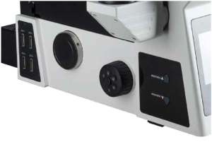
All motorized Control System.
The Z-axis of BCF297 laser confocal microscope adopts a motorized device, which can quickly adjust the Z-axis height according to the real-time image. AF one-key autofocus, eliminating the need for fine-tuning steps and improving work efficiency. Integrated control buttons on both sides of the frame, can quickly switch or rotate the condenser, brightness, objective lens, attenuator disc and fluorescence disc, to improve the operation convenience.
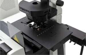
High-speed, high-precision stage.
The stage ensures exceptional stability and precise positioning for detailed sample control during observation. It enables fast and smooth scanning, significantly enhancing data acquisition efficiency and image quality. It’s the ideal choice for scientific research requiring both efficiency and precision.
Front LCD panel
It is able to display the status of electric parts in real time, and set the observation mode, switch the light brake, etc. It greatly improves the user experience and makes the research work more convenient.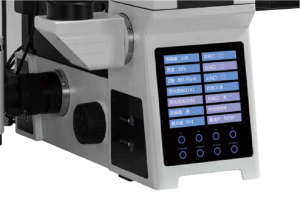
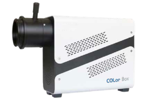
Multiband LED fluorescence illumination system
Equipped with multiple excitation channels for quick switching, this system adapts perfectly to different experimental needs. The advanced light intensity control technology ensures optimal illumination every time. Coupled with its long-lasting durability and intelligent SDK control, it takes your scientific exploration further. Additionally, it supports live-cell observation expansion.
High scalable
The big frame provides sufficient space for third-party configurations. Single-layer optical path or double-layer optical path can be selected as required. It can be loaded into 16 filters at most, providing maximum scalability for the in-depth research. It supports live-cell observation expansion.
Distinguished Confocal Imaging Optical Components
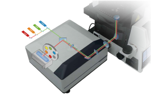
Innovative confocal pinhole unit
Pinhole design is based on the principle of light reversibility. The excitation light of the lamp and the emission light of the sample pass through the same pinhole, and they keep a 100% conjugate relationship. It not only ensures the acquisition efficiency of fluorescence signal, but also improves the filtering of non-focal plane signal, for the higher detection sensitivity and better image resolution.
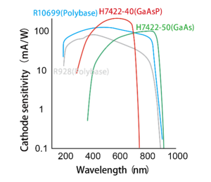
Controller detection unit
The new generation of highly sensitive GaAsP technology enables automated multicolor confocal fluorescence imaging, allowing researchers to perform detailed analysis of complex samples more easily and quickly.
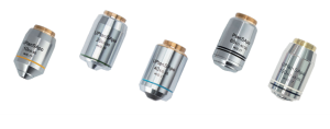
SAPO series super apochromatic objectives
Converging the optic axises of red, green and blue to one focal plane, correcting the axial chromatic aberration of violet light, the original color of samples such as red/green/blue is able to be presented. The resolution and brightness are improved based on large numerical aperture. Wide spectrum correction ensures flat field and increase the color difference correction range from 400nm to 1000nm.

SAPO 60X transmittance curve
Special software for laser confocal
This system is equipped with high-sensitivity, high-resolution, and high-efficiency imaging technology, meeting the diverse needs of complex data acquisition. The software supports multichannel acquisition, Z-stack imaging, and large fluorescence image stitching, providing users with a comprehensive imaging solution. For image processing, the software integrates functions such as image fusion, image enhancement, 3D reconstruction and dynamic image generation, significantly improving image presentation quality. In terms of image analysis, the software offers advanced features like optical density analysis and colocalization analysis, delivering a professional and powerful user experience.
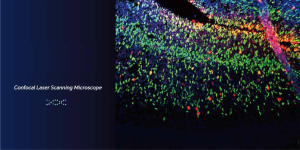
Image acquisition
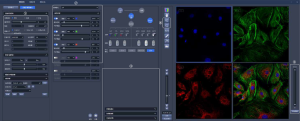
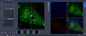
① This module allows users to freely adjust confocal scanning parameters, including scanning speed, image resolution, magnification, laser power and gain, to suit different samples and experimental needs.
② Achieve six-dimensional (x, y, z, λ, t) imaging, capturing comprehensive sample information.
Provide various custom acquisition modes, such as xyz, xyt, xyzt, xyλ, xyλt, xyzλt, to meet different experimental requirements.
Support real-time stitching of large images, seamlessly integrating multiple fields of view for a complete view.
Enhance image depth perception through depth of field extension technology.
Apply pseudo-color marking in real-time to improve the visual effect and interpretability of images.
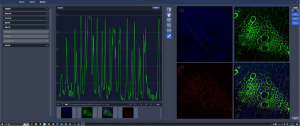
③ Support real-time optical density measurement for analyzing the transparency characteristics of samples. Provide real-time histogram display to understand the statistical distribution of images instantly.
④ Allow users to zoom in on images for detailed observation or a general overview.
⑤ Intelligently adjust histogram thresholds to achieve optimal image contrast.
Image processing
Support advanced 3D reconstruction and visualization, bringing three-dimensional structures to life.
Ensure accurate overlay of multi-channel images through precise colocalization processing.
Synchronize analysis of multiple marker points through colocalization linkage.
Provide diverse image editing tools, including flipping, mirroring, and background removal.
Generate dynamic image sequences to capture biological events in the time dimension.
Stack processing function supports three-dimensional reconstruction and time series analysis.
ROI (Region of Interest) processing focuses on detailed analysis of specific areas.
Image analysis
Provide a series of morphological parameter analyses, including perimeter, area, roundness, etc.
Measure the highest and lowest grayscale values of images to evaluate the contrast range.
Perform colocalization analysis to quantify spatial relationships between multiple markers
Count cells and particles for various quantitative analysis experiments.
Application
- Biological and life sciences
Cell biology
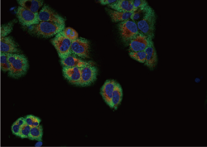
BCF297 is able to accurately image all cells labeled with various fluorescent proteins and multi color probes, studying the fluorescence colocalization, dynamic properties and spatial relationships of two or more target proteins. Besides, BCF297 can achieve the morphological structure of 3D cell culture such as organoids/globules by 3D reconstruction, finding out more hidden information.
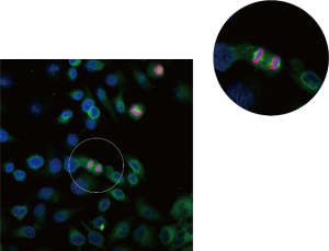
HELA Cell (Peking University Health Center) 60X
Human hepatocytes (Zhejiang University) 40X
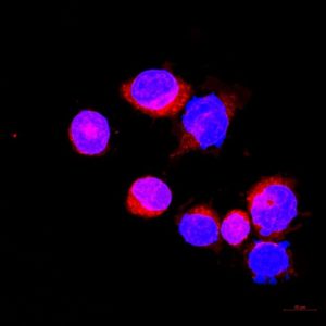
Biochemistry and molecular biology
BCF297 is a powerful tool for studying molecular and protein interactions, intracellular signaling pathways, and gene expression. It provides precise quantitative data on the exact locations and dynamic processes of these biomolecules within cells.
Published in: <The Journal of Nutritional Biochemistry>.
Image source: Department of Child and Adolescent Health, Public Health College, Harbin Medical University
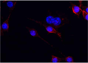
Iba-1 Cells (Harbin Medical University)
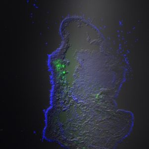
Development biology
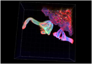
BCF297 is ideal for observing model organisms, such as zebrafish and fruit flies, which have large, complex and dense structures. BCF297 is suitable for non-destructive imaging, with its wide-field imaging and layer scanning functions helping to obtain detailed structural images of samples and present details from different depths of the sample, aiding development and growth research.
Dicotyledon stem (Huazhong Agricultural University) 60X
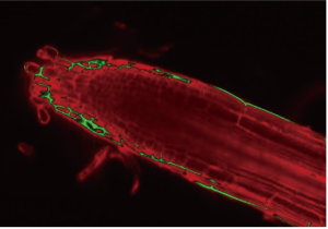
Arabidopsis root (Huazhong Agricultural University) 40X
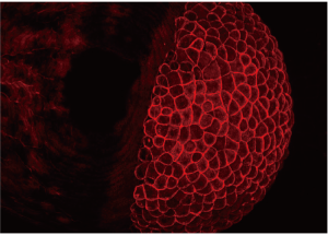
Zebrafish embryo (West China Hospital) 20X
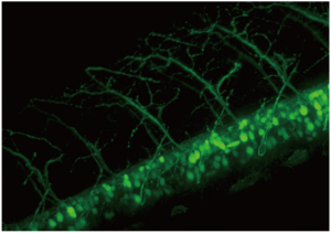
Zebrafish nerves (West China Hospital) 40X
Materials science
Biomaterial
In the field of bio-photonics, the research heat of biological information and nanomaterials is increasing. In the study of photoelectric materials, BCF297 with the living cell environment monitoring can cooperate with the living cell environment monitoring module to observe the process of interaction between materials and cells for the fusion of new functional materials, inorganic nano hybrid materials and living cells.
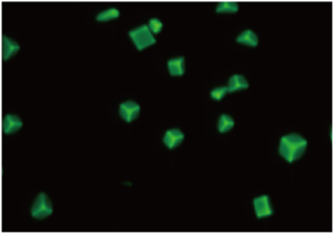
Functional materials (Wuhan University of Technology) 60X

293T cells (Wuhan Polytechnic University)
Published in: Colloids and Surfaces B: Bio interfaces
Image source: Chen Xin Research Group, Wuhan Polytechnic University
Medical and pharmacological
Pharmaceutical Chemistry
Can be used to study the mechanisms of drug actions in cells and tissues and screen for new drug candidates.
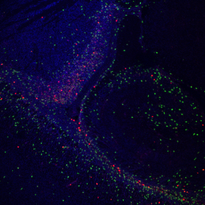
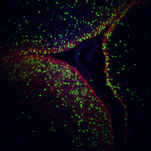
Pathology
The layer scanning of BCF297 is suitable for different histopathological sections of animals and plants, especially for large tissue. Much more details and more accurate data are available.
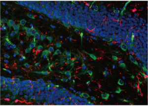
Hippocampal slice (Chinese Military Academy of Science) 40X
Agricultural, forestry and food science
Food safety and quality control
Utilizing its advanced imaging technology, BCF297 provides excellent resolution and image clarity, effectively reducing background signal interference and enhancing image contrast. This ensures clear observation even with complex or opaque samples such as high internal phase emulsions (HIPEs). The brightness and penetration of its laser light source offer significant advantages for in-depth analysis and understanding of these challenging samples.
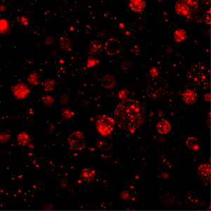
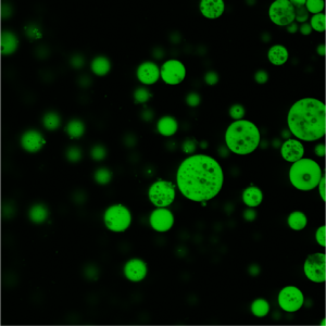
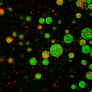
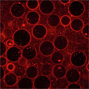
High Internal Phase Emulsions (HIPEs) (Xihua University)
Published in: Food Hydrocolloids
Published in: Food Research International
Image source: Professor Chen Xianggui’s Research Group
School of Food Science and Engineering, Xihua University
Specification
|
Item |
Specification |
||
|
Laser |
405nm, 488nm, 561nm, 640nm |
||
|
Scan Unit |
High speed optical scanning galvanometer |
||
|
Field of view: (≥20) |
|||
|
Scanning element: 512*512-8192*8192 |
|||
|
Pixel time: 0.5μs-10μs |
|||
|
Standard scanning speed: 3fps (512*512) |
|||
|
Scanning: 1X-50X |
|||
|
Pinhole |
35/45/55/65/85/105μm |
||
|
Probe Unit |
Multi alkali PMT, QE≥25%@500nm |
||
|
GaAsP PMT, QE≥45%@500nm |
|||
|
Optical System |
Infinity Color Correction Optical System |
||
|
Viewing tube |
20-45° tilting binocular head, inverted image |
||
|
Eyepiece |
High eyepoint wide field plan eyepiece PL10X/22mm, diopter adjustable, micrometer attachable |
||
|
Microscope Body |
Low position coarse and fine coaxial electric focusing mechanism, range 10.5mm, precision: 0.01μm |
||
|
Stage |
Stage size: 350mm (X)*200mm (Y), moving range: 114mm (X)*75mm (Y), maximum speed: 50mm/s, with slide holder and Φ36 petri dish holder. |
||
|
Objectives |
Infinity Plan Super Apochromatic Objectives |
10X/NA=0.4, WD=3.1mm |
|
|
20X/NA=0.8, WD=0.6mm |
|||
|
40X/NA=0.95, WD=0.18mm |
|||
|
(Oil) 60X/NA=1.42, WD=0.17mm |
|||
|
(Oil) 100X/NA=1.45, WD=0.13mm |
|||
|
Condenser |
Electric seven-hole condenser, NA=0.55 |
||
|
Transmitted Illumination |
10W LED transmitted illumination |
||
|
Fluorescent Module |
Fluorescent illumination, 8-hold fluorescence rotating system, supporting up to 16 wells |
Multi-band LED light source module, 10W high- power LED |
|
|
Fluorescent mercury lamp light source module, 100W DC mercury lamp |
|||
|
DIC Imaging |
DIC detector |
||
|
Camera |
5MP/20MP camera |
||
|
Operating Software |
Image acquisition, image processing, image analysis, including functions such as large image stitching, 3D reconstruction and cell counting. |
||
Note: ● Standard Outfit, ○ Optional
Sample Images


Dimension

Unit: mm

 LED Research Fluorescent Biological Microscope")


