


BDEM-18 Desktop Tungsten Filament Scanning Electron Microscope
Introduction
With outstanding performance, high-speed imaging, diverse signals, BDEM-18 desktop scanning electron microscope signal acquisition bandwidth up to 10M, fast scanning speed, video mode real-time observation of samples, no ghosting, dragging, do not miss every detail. Compatible with a variety of in-situ functional sample stage.
Details
Overview
Packaging & Delivery
Packaging Details:Strong Carton with Polyfoam Protection
Port:Beijing
Lead Time:Within 2-4 Weeks after Receiving Payment
Introduction
Introduction
With outstanding performance, high-speed imaging, diverse signals, BDEM-18 desktop scanning electron microscope signal acquisition bandwidth up to 10M, fast scanning speed, video mode real-time observation of samples, no ghosting, dragging, do not miss every detail. Compatible with a variety of in-situ functional sample stage.
Features
1.All operation can be done with just the mouse.
2.Highly integrated optoelectronic design eliminates the need for manual hardware configuration.
3.Optional 10kV deceleration sample stage.
4.Vacuuming time less than 90s.
5.Fast scanning speed real-time observation of samples in video mode, so as not to miss every detail.
6.SE detector, BSE detector, EDS and many other detectors. (SE+BSE mode simultaneous acquisition, arbitrary superposition.)
7.Continuously adjustable accelerating voltage, adapting to many types of samples and in-situ testing functions.
8.No need to install additional shock absorbers, plug and play with regular utility power.
9.Rich in-situ expansion function, compatible with stretching sample stage, heating stage, TEC cooling stage, etc.
Specification
|
Item |
Specification |
|
Environmental Requirements |
AC 220V, 50Hz, 1kW. No shock absorber required |
|
Acceleration Voltage |
3kV~18kV continuously adjustable, 1kV step |
|
Electron Gun |
Pre-centered tungsten filament, integrated condenser lens, no need to manually adjust the objective diaphragm |
|
Magnification |
200,000X |
|
Detector |
Four-division backscattered electron detector |
|
|
Secondary electron detector |
|
|
Integrated spectrometer (optional) |
|
Stroke of Sample Stage |
X/Y motorized sample stage, moving stroke: 30mm*30mm |
|
Maximum Sample Size |
Ф50mm*35mm (H) |
|
Working Distance |
5-35mm |
|
Vacuum Mode |
High vacuum mode: vacuum extraction time less than 90s Low vacuum mode (optional): 1-60Pa automatic control |
|
Imaging Mode |
Video Mode: 512*512 pixels without small window scanning. Fast Scan Mode: 512*512 pixels. Slow Scan Mode: 2048*2048 pixels. Image Format: BMP, TIFF, JPEG, PNG |
|
Navigation Function |
Optical CCD navigation |
|
Automatic Function |
One-touch auto-configuration of brightness, contrast, auto-focus, etc. |
|
Sizes |
Main unit: 283mm*553mm*505mm Mechanical pump: 340mm*160mm*140mm |
|
Extended Function |
Compatible with a wide range of in-situ functional sample stations (tensile, heated, TEC, and other in-situ test systems) |
Note: ● Standard Outfit, ○ Optional
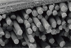
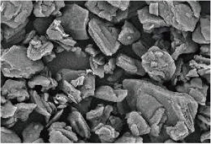
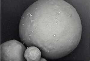
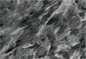
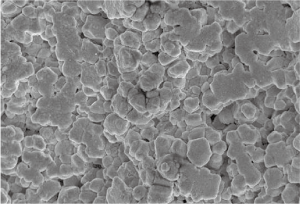
Sample Images
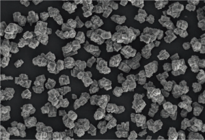
Particle detection
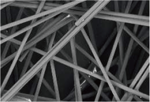
Fiber testing
Industrial monitoring
Anode materials for lithium battery
Powered medicine material
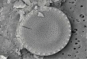
Diatom
Hydrogel
Aluminum nitride
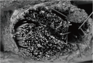
Tubular nanomaterials
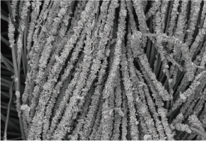
Functional nanomaterials
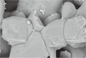
Functional inorganic material
Research and teaching


