What is a Compound Microscope?
 Optical microscopes are crucial tools in scientific research, medical service, and education.
Depending on differences in observation methods, applications, and operating principles, microscopes can be classified into various types. This article will start from the working principles of microscopes to help you better understand compound microscopes.
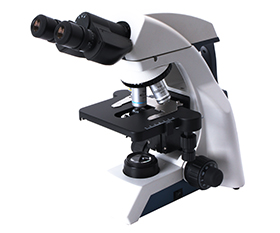
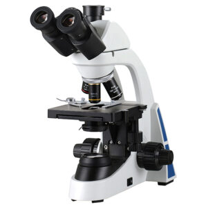
The compound microscope is a high-magnification microscope that uses multiple lenses to magnify and observe specimens. It consists of objective lenses and eyepieces, with the magnification of the objectives typically being 4x, 10x, 40x, and 100x, while the eyepiece magnification is usually 10x. Transmitted light passes through the specimen, is focused by the objective lenses, and then passes through the eyepiece, allowing you to see a clear two-dimensional image.
What are the Parts of a Compound Microscope and Their Functions?
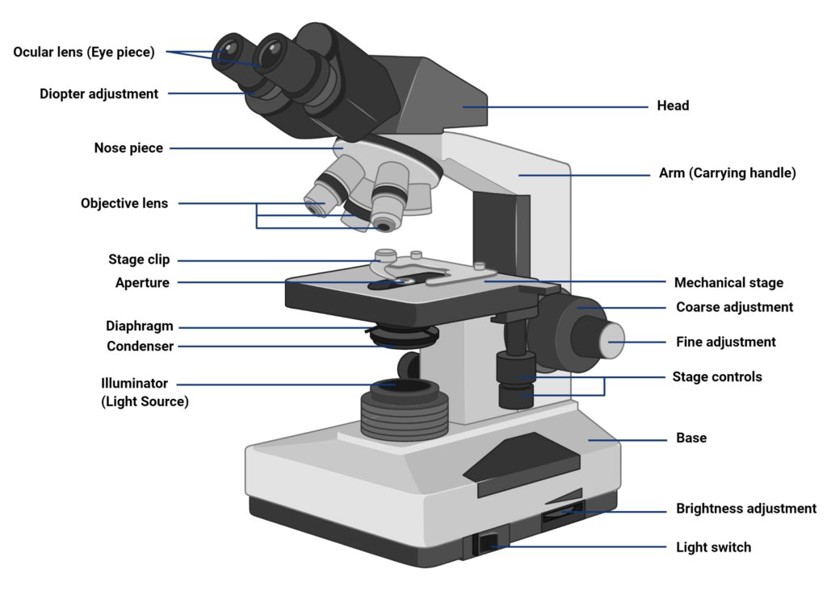
Objective Lens:
The objective lens is a crucial part of a compound microscope, made up of multiple lenses forming a lens system. Its job is to magnify the specimens at different levels. It’s important to note that the 100x objective lens is a high-magnification lens that requires immersion oil for use.
Eyepiece:
The eyepiece is at the top of the microscope and its role is to further magnify the image produced by the objective lens, allowing you to achieve the final level of magnification.
Light Source:
The light source is used to illuminate the specimen and can be categorized into transmitted light and reflected light. Common types of light sources include halogen, mercury lamps, and LED. The advantages of LED, such as low power consumption and long lifespan, have made it increasingly popular in light source selection.
Condenser:
The condenser is another important optical component, consisting of one or more lenses. It is located beneath the stage and often comes with an adjustable aperture. The light source is focused onto the specimen through the condenser, and its role is to regulate and control the light source, ensuring even illumination.
Stage:
The stage is a platform used to hold the specimen. The specimen is secured in place using stage clips, and the stage is typically movable in the X-Y directions. Adjustments for the specimen’s position can be made manually or automatically, allowing both horizontal and vertical movement.
Nosepiece:
The nosepiece is used to secure the objective lens and switch magnification levels.
Focusing:
Focusing involves both coarse and fine adjustments. Coarse focusing quickly moves the objective lens to find the image’s rough focus, while fine focusing involves small, precise adjustments to achieve a clearer image.
Base:
Used to keep the microscope stable.
What are the Applications of Compound Microscopes?
Cellular, Pathological, and Microbial Research
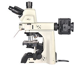
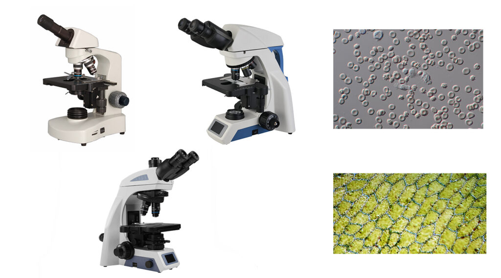
The biological microscope is the most common type of compound microscope used to observe biological specimens at high magnification. Phase contrast microscope is a special type of biological microscope that differs from the ordinary brightfield microscope. It requires phase contrast objective lenses and a condenser to alter the phase of light passing through the specimen, thereby enhancing contrast. It is mainly used for observing blood cells, live cells, and unstained transparent specimens.
The fluorescence microscope is used to observe specimens with natural fluorescence or those stained with fluorescent dyes. This technique allows the labeling of different cellular structures, such as mitochondria, cell nuclei, and cell membranes. Fluorescence microscopy is widely applied in fluorescence in situ hybridization (FISH) techniques for analyzing and detecting the distribution and localization of nucleic acids.

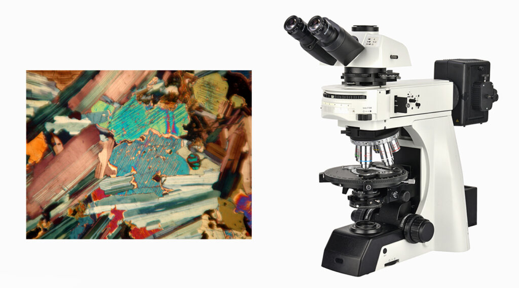
Mineral and Rock Analysis
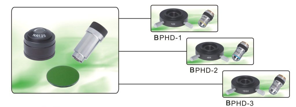
The polarizing microscope is another common type of compound microscope, featuring two crucial polarizing devices – the polarizer and the analyzer. The polarizer is positioned between the light source and the specimen, converting natural light into polarized light. The analyzer is located between the specimen and the eyepiece and is used to analyze the vibration direction of light passing through the specimen. The polarizing microscope is commonly used for the analysis of the structure and properties of minerals and rocks.
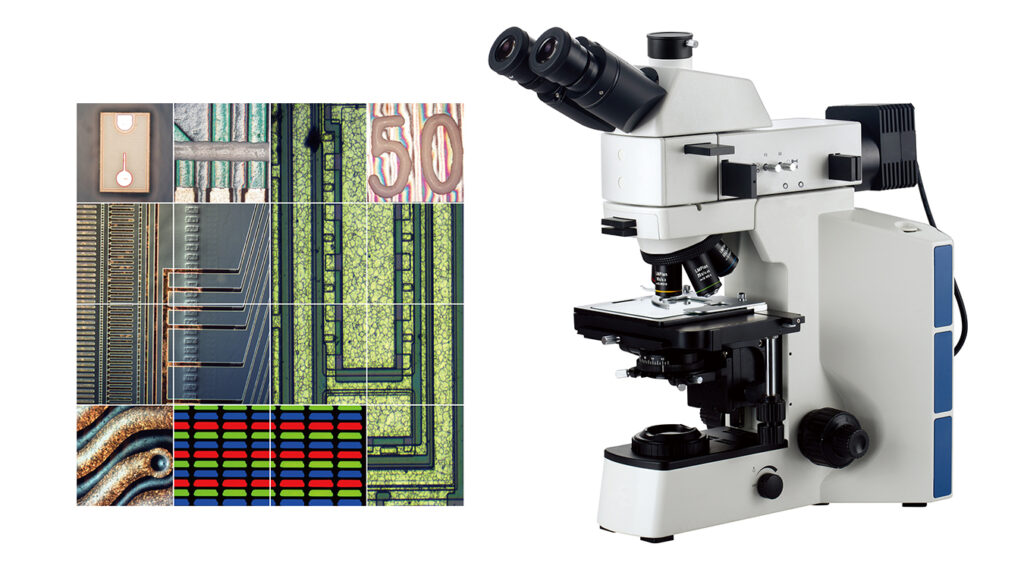
Metal Analysis
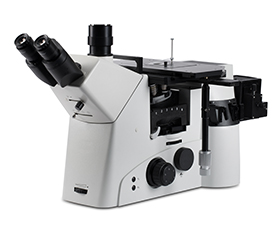
The metallographic microscope, or metallurgical microscope, is also a type of compound microscope primarily used for observing metals, alloy materials, semiconductor testing, ceramics, rocks, and other opaque materials. Therefore, metallographic microscopes typically employ high-intensity reflected light, which is directed through the objective lens onto the specimen, producing high-contrast images of opaque specimens. They can also simultaneously utilize both transmitted and reflected light.
Compound Microscopes VS Stereo Microscopes
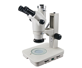
The stereo microscope, also known as a low-magnification microscope or dissecting microscope, is another common optical microscope. It features two independent optical paths, each leading to a separate eyepiece. This design allows observers to see a true 3D stereoscopic image. No prepared slides are needed for observation, and the microscope typically provides a long working distance. It is commonly used for observing larger, opaque specimens such as circuit boards, gemstones, insects, and other objects.
|
Compound Microscope |
Stereo Microscope |
|
|
Magnification |
Higher Magnification Range 40x-1000x |
Lower Magnification Range 6x-50x |
|
Field of View and Depth of Field |
Small observation field, presenting a flat image. |
Broad observation field, presenting a true 3D stereoscopic image. |
|
Working Distance |
Short working distance, requiring the objective lens to be close to the specimen to focus and obtain a clear image, especially when observing tiny transparent specimens. |
Longer working distance, providing more significant operating space and facilitating adjustments and movement when observing larger specimens. |
|
Applications |
Biology, histology, pathology, bacteriology, immunology, and observation of tiny objects.
Identification and analysis of metals and alloys.
Analysis and research in geology, minerals, and materials science. |
PCB, SMT, electronic device testing, semiconductor chip inspection, metal and material testing, precision part testing.
Coin collecting, gemology, gemstone inlaying, engraving.
Maintenance and inspection of small parts. |
Advantages and Disadvantages of Compound Microscopes
In summary:
The Advantages of a Compound Microscope:
High magnification, allowing the observation of smaller objects.
Versatile applications, suitable for observing various types of specimens.
The Disadvantages of a Compound Microscope:
It has only one optical path, resulting in a smaller field of view and limited depth of field.
It cannot observe the deep-level structures of specimens.
It requires higher standards for specimen preparation.
If you have any questions about choosing a microscope, please feel free to contact us. BestScope provides you with professional solutions.