


BCM2-300 Live Cell Imaging System
Introduction
The BCM2-300 series is a fully automatic live cell imaging system, with bright field, phase contrast and 3-channel fluorescent observation. It abandons the bulky housing and complicated operating steps of ordinary biological microscopes, allowing cell observation in one step. The built-in high-speed and high-sensitivity camera has a time-lapse shooting function, which can fully record the cell culture process.
Details
Overview
Packaging & Delivery
Packaging Details:Strong Carton with Polyfoam Protection
Port:Beijing
Lead Time:Within 2-4 Weeks after Receiving Payment
Introduction
The BCM2-300 series is a fully automatic live cell imaging system, with bright field, phase contrast and 3-channel fluorescent observation. It abandons the bulky housing and complicated operating steps of ordinary biological microscopes, allowing cell observation in one step. The built-in high-speed and high-sensitivity camera has a time-lapse shooting function, which can fully record the cell culture process.
Features
1. Bright field/ phase contrast
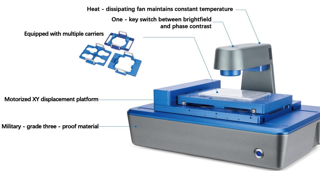
One-key switch between bright field and phase contrast for observing different samples.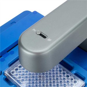
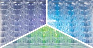
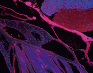
2. 3-channel fluorescence
Standard with 3 color excitation band fluorescence, B/G/U, motorized one-key switch.
3. Sealed, waterproof and corrosion resistant
Sterilizable via alcohol, double-distilled water/pyrroloquinoline, UV.
Usable in CO2 incubators.
4. Compatible with most common carriers
Compatible with multiple carriers, fitting various cell plates, dishes, flasks, slides and other containers, suitable for diverse experiments.

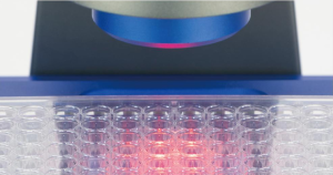
5. Low phototoxicity LED light source
625nm red LED bright field illumination, with low photobleaching and cytotoxicity, to protect samples and extend cell observation time.
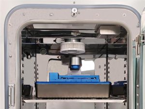
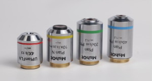
6. Motorized dual objectives
Pre-set diaphragm with dual-objective configuration, optional magnifications of 4X/10X/20X objectives.
7. Motorized X/Y stage & Z-axis focus
Motorized stage with X/Y scanning and motorized Z-axis, with auto-intelligent focusing.
8. One-click whole well scan and stitching
Navigational scanning settings enable grouping, free-arm skipping of any regions, stitching of scans of adjacent fields of view and stitching of scans of the entire field of view.


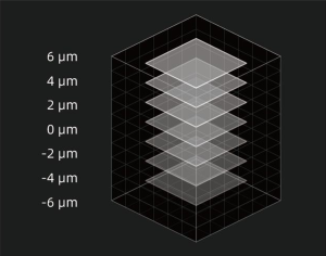
9. Z-stack scan
High-precision motorized Z-axis: freely set Z-stack range/step for automatic scanning and layer imaging. Defocus removal algorithms enable clear imaging of thick samples, e.g., organoids.
10. Multi-channel scan & overlay
Navigational settings for multi-channel scanning, including multi-color fluorescence as well as bright field and phase contrast channels, enabling overlay output of images from any two or more channels.

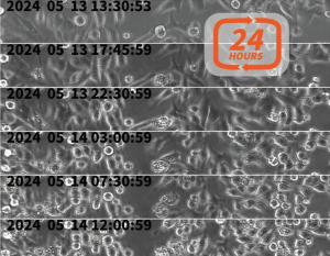
11. Continuous time lapse acquisition
Long-term continuous shooting can generate time-lapse videos of any field of view and any duration, presenting complete information of the whole cycle of living cell samples.
12. Intelligent analysis functions
Comes with image recognition and AI deep learning modules. Matches optimal analysis algorithms for different cell experiments, expandable with advanced modules like organoid, nerve cell and tumor sphere analysis.

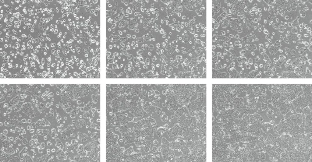
13. Cell confluency
Cell confluency is the percentage of surface area covered by cells in 2D culture. Timed live-cell imaging and data analysis determine confluency, plotting confluency-over-time curves and cell-count curves per channel. It is widely used in cell passaging, transfection, virus infection, drug treatment, etc.
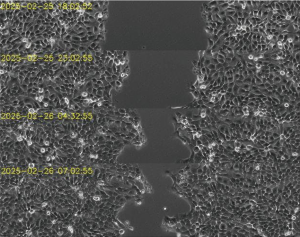
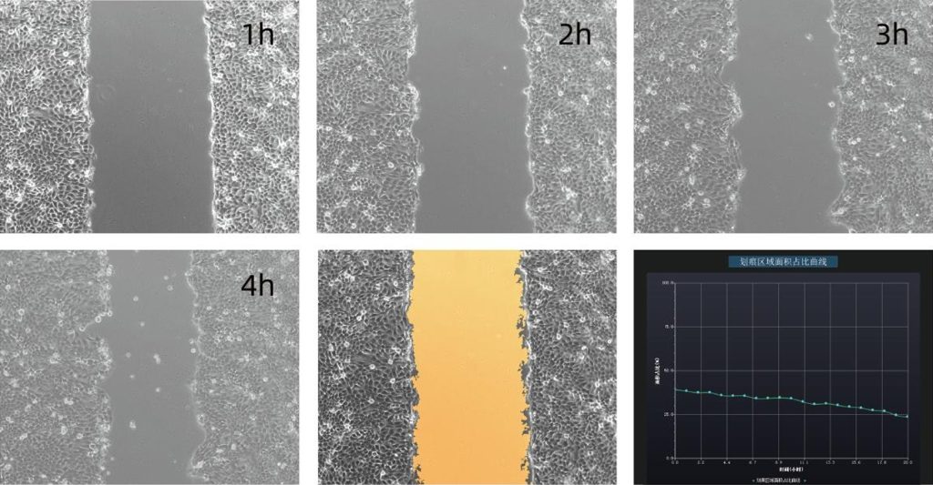
14. Scratch assay
The cell scratch assay evaluates adherent cell migration. Create a scratch on the dish to simulate a wound, imaging at preset time points and quantitatively measuring cell migration between two rows. It is used for researching cell proliferation, migration, wound healing and drug screening.
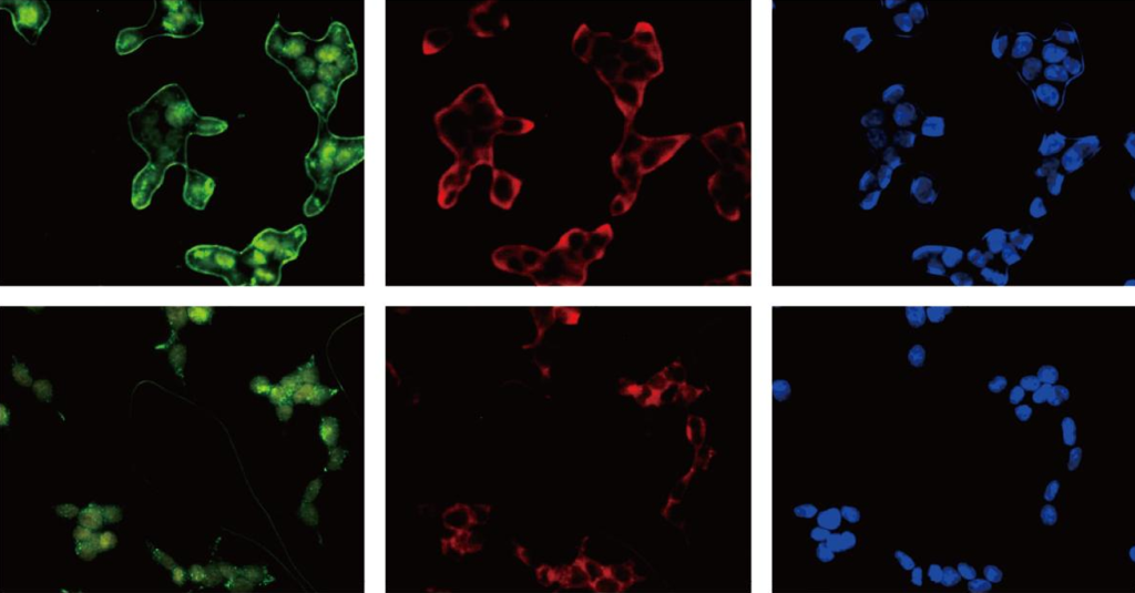
15. Fluorescence transfection efficiency
Transfected cells with fluorescent label are imaged at different time points post-transfection. Cell counting function counts and record cells per channel, used in gene research, drug development, CGT, stem cell and organoid studies, etc.
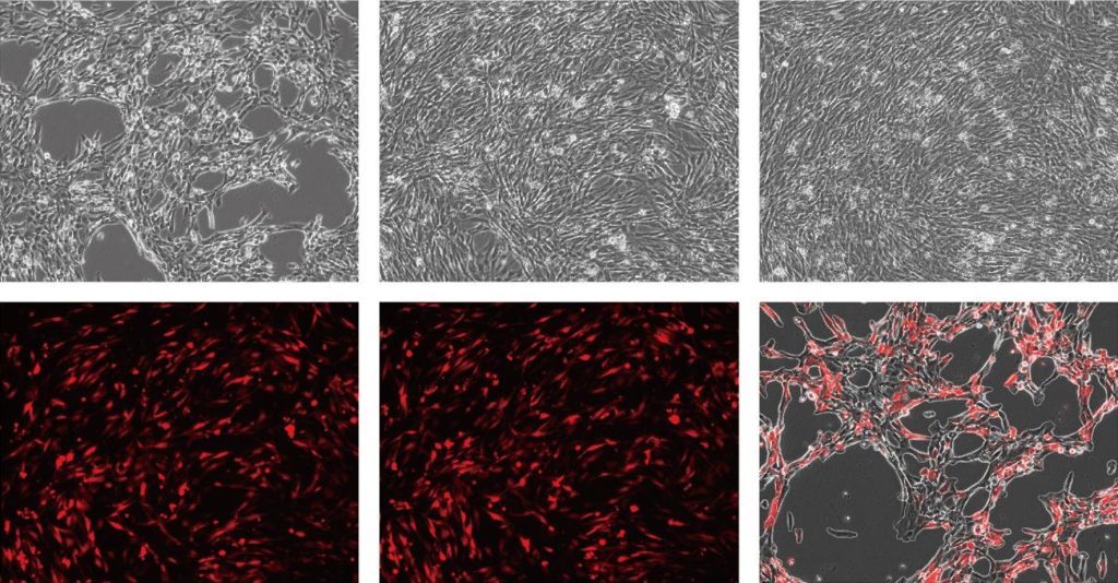
16. Cell viability
Bright field and fluorescence images are taken. Cell counting counts total and dead cells (fluorescent). Time-point data records generate a cell viability curve. It is used in cell toxicity, drug development, CGT, vaccine R&D and production.
Application
1. Dynamic monitoring of organoids
As 3D culture models, organoids fill the gap between 2D cells and animal models, widely used in disease simulation, drug testing and regenerative medicine.
Kinetics of organoid formation
Quantitatively monitor the impact of seeding density on formation efficiency. Track the aggregation process and record the tensor of area/volume changes.
Migration ability assessment
Invasive analysis of tumor organoids.
Drug sensitivity testing
Real-time recording of the morphological collapse of organoids in response to chemotherapy drugs.
2. Drug development
Live-cell imaging replaces traditional endpoint methods, enabling dynamic quantification of drug effects and accelerating preclinical research.
Internalization and bystander effect of ADC drugs
Dual-color fluorescence tracking of the intracellular transport of antibody-drug conjugates.
Dynamic toxicity assessment of small molecules
Killing efficacy of immune cells
Recording the lysis process of tumor spheroids by NK cells.
3. Development of stem cell therapy products
The activity and differentiation stability of stem cells are the core of quality control for therapeutic agents and require non-destructive long-term monitoring.
Quality control of proliferation activity
Auto-confluency analysis of umbilical cord MSCs for harvest remainders.
Differentiation tendency early warning
Tracking early morphological changes in adipogenic/osteogenic differentiation.
Stability of cell therapeutic products
Verification of survival rate after cryopreservation and recovery.
4. Monitoring of cell industrial processes
Monitor cell culture conditions to evaluate culture status, experimental status, and subculture timing and control quality.
Batch-to-batch consistency management
Comparison of proliferation curves of CHO cells from different batches.
Real-time contamination early warning
AI-based morphological recognition of bacterial/fungal contamination.
Stability of cell therapy products
Monitoring of aggregation behavior during CAR-T cell expansion.
5. Agricultural science
Plant-pathogen interaction research, fertility and organelle localization of beans, gene function and protein expression research, etc.
6. Cell industry
Cell culture experiment monitoring, in-line quality control of bioreactors, stability monitoring of cell therapy products, etc.
7. Immunology
Macrophage phagocytosis tracking, neutrophil activation analysis, collagen-induced immune monitor, etc.
8. Neuroscience
Neuron network formation and construction, research on neural stem cell differentiation, quantitative analysis of brain organoids, etc.
9. Drug R&D
Preliminary toxicity screening of chemical drugs, early activity evaluation of compounds, research on drug action mechanisms, etc.
10. Regenerative medicine
Skin wound healing model, basic research on adipogenesis, monitoring of adhesion behavior of tissue engineering scaffolds, etc.
11. Stem cell applications
Pluripotency detection of stem cells, early warning of directed differentiation, dynamic monitoring of organoid morphology, etc.
12. Tumor research
Detection of apoptosis and necrosis, analysis of tumor cell migration and invasion, evaluation of chemotherapeutic drug activity, etc.
13. Aquaculture
Fish health assessment and germplasm optimization, fish immune invasion early warning, reproductive cell viability analysis, etc.
Specification
|
Item |
Specification |
|
Bright Field Observation |
Transmitted phase contrast illumination, working distance 55mm |
|
50,000 hours long lifespan, 625nm low-phototoxicity LED light source |
|
|
Fluorescence Observation (Fluorescence Wavelengths Customizable) |
Blue (B): 472-495nm |
|
Green (G): 543-560nm |
|
|
Ultraviolet (UV): 393-416nm |
|
|
Objective |
Select any two from 4X/10X/20X objectives |
|
Motorized objective switching |
|
|
Stage |
Motorized X/Y moving stage, moving range: 115mm*77mm, repeatability accuracy ≤3μm |
|
Focusing System |
Motorized, auto-focus mechanism |
|
Camera |
High-speed and high-sensitivity camera, 5MP, 2/3”, 40fps |
|
Software |
Supporting remote control, including illumination control, camera control, photo taking, video recording, time-lapse photography |
|
Analysis Function |
Cell counting, cell confluency, scratch assay, transfection efficiency, viability analysis |
|
Scanning Function |
Multi-well imaging: custom-set X/Y array for multi-point photography, bright field and fluorescence multi-channel overlay |
|
Region scanning: set rectangular regions for full-scan stitching, bright field and fluorescence multi-channel overlay |
|
|
Data transmission / Power Supply |
USB data cable, DC power Cable |
|
Working Environment |
5-40°C, 20-95% RH |
|
Dimensions |
252mm*377mm*208mm |
|
Package Dimension & Weight |
55cm*43cm*35cm, 14.1kg |
Dimension
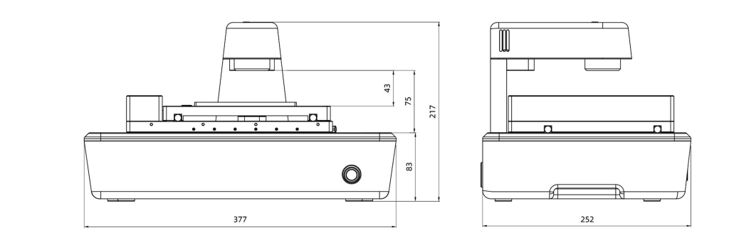
Unit: mm


