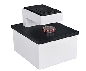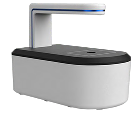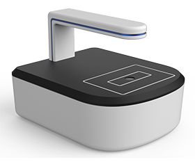

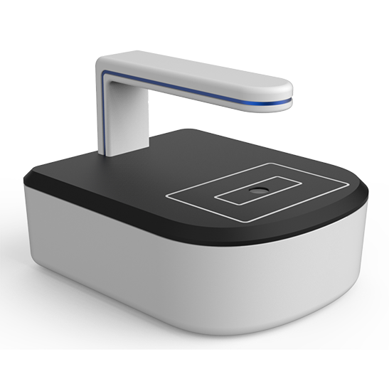
BCM2-200A Live Cell Imaging System
Introduction
Compatible with automated bright field imaging in incubator, BCM2-200 Live Cell imaging System allows for long-term dynamic monitoring and analysis of live cells. With easy-to-use software and an Al intelligent analysis system, it can timely help you visualize and analyze sample morphology and behavior changes, and can also realize cell culture progress reminders.
Details
Overview
Packaging & Delivery
Packaging Details:Strong Carton with Polyfoam Protection
Port:Beijing
Lead Time:Within 2-4 Weeks after Receiving Payment
Introduction
Introduction
Compatible with automated bright field imaging in incubator, BCM2-200 Live Cell imaging System allows for long-term dynamic monitoring and analysis of live cells. With easy-to-use software and an Al intelligent analysis system, it can timely help you visualize and analyze sample morphology and behavior changes, and can also realize cell culture progress reminders.
Features
1.Simple workflow
BCM2-200 Live Cell Imaging System greatly simplifies your workflow through automated scheduled observation, analysis and reporting, you can remotely monitor the progress of cell culture without entering the clean room or opening the incubator, greatly improving work efficiency and avoiding the risk of disturbing growth and contaminating samples.
2.Phase contrast and fluorescence imaging
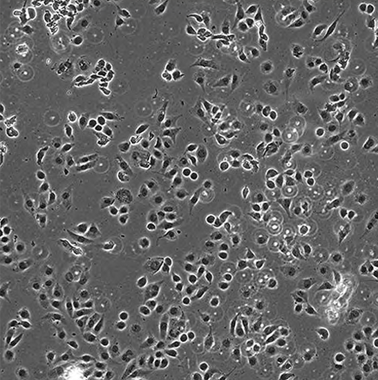
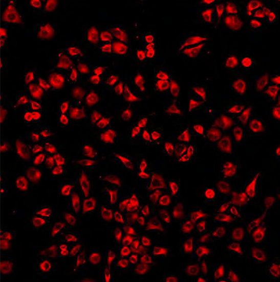
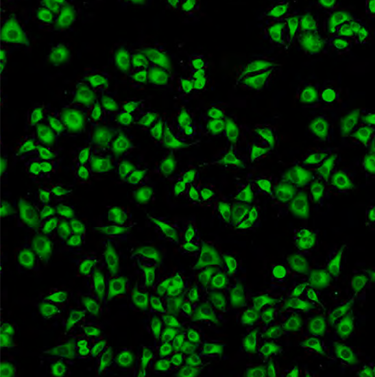
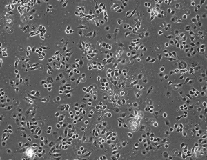
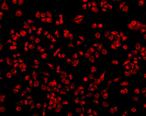
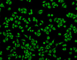
Combine bright field, phase contrast and fluorescence imaging, equipped with blue and green fluorescence channels, suitable for monitoring cell transfection and cell viability scenes. It provides high-quality phase contrast and fluorescence imaging.
3.Software
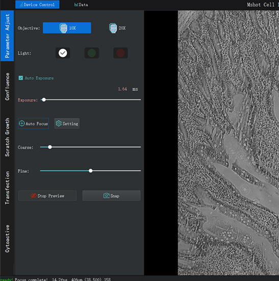
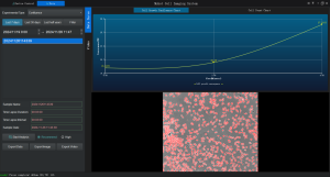
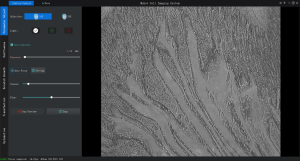
It supports confluence analysis, cell counting, time-lapse recording, email reminder, remote control, etc.
Specification
|
Item |
Specification |
BCM2-200A |
BCM2-200B |
|
Bright Field Observation |
Transmitted phase contrast illumination, working distance 70mm |
● |
● |
|
Long life low phototoxicity LED light source, 625nm |
● |
● |
|
|
Fluorescence Observation |
Blue (B): 475/30nm |
● |
● |
|
Green (G): 560/40nm |
● |
● |
|
|
Three colors of fluorescence are available, and the wavelength can be customized |
○ |
○ |
|
|
Objective |
Single objective, 4X/10X/20X optional |
● |
|
|
Dual objectives, motorized switching, select any two from 4X/10X/20X objectives |
|
● |
|
|
Stage |
Manual moving stage |
● |
● |
|
Focusing System |
Motorized, auto-focus mechanism |
● |
● |
|
Camera |
High-speed and high-sensitivity fluorescence camera, 5MP, 2/3”, 40fps |
● |
● |
|
Software |
Adopts C/S architecture and support remote control, including lighting control, camera recording photography, video recording, time-lapse photography, cell counting, cell confluency, scratch experiment, external network control, email reminder |
● |
● |
|
Data transmission / Power Supply |
USB data cable, DC power Cable |
● |
● |
|
Working Environment |
5-42°C, 5-95% RH |
● |
● |
|
Dimensions |
214mm*260mm*182mm |
● |
● |
Note: ● Standard Outfit, ○ Optional
Dimension
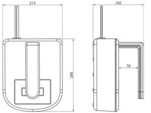
Unit: mm