


BCM3-300F3 Live Cell Imaging System
Introduction
The BCM3-300 Live Cell Imaging System is a scientific research instrument that can monitor the growth status of cells in real time in a carbon dioxide incubator. It can conduct high-throughput live cell growth monitoring in real time and support video replay and data analysis. It can greatly reduce manual operation and lower the risk of cell contamination. While keeping track of cell dynamics in real time, it will not interfere with cell growth. It is perfectly suitable for most cell growth research.
Details
Overview
Packaging & Delivery
Packaging Details:Strong Carton with Polyfoam Protection
Port:Beijing
Lead Time:Within 2-4 Weeks after Receiving Payment
Introduction
Introduction
The BCM3-300 Live Cell Imaging System is a scientific research instrument that can monitor the growth status of cells in real time in a carbon dioxide incubator. It can conduct high-throughput live cell growth monitoring in real time and support video replay and data analysis. It can greatly reduce manual operation and lower the risk of cell contamination. While keeping track of cell dynamics in real time, it will not interfere with cell growth. It is perfectly suitable for most cell growth research.
Features
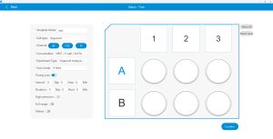
1.User-friendly interface
The program is easy to set up. The users can freely shoot or preset the shooting process. It supports single-field, 4-field and 9-field shooting for each hole.

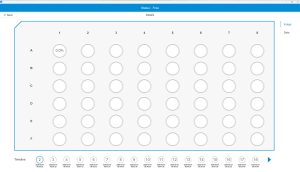
2.Intelligent automation
BCM3-300 supports fully automatic high-throughput scanning and Imaging. Historical templates can also be called up.
3.Low power consumption and high throughput
The device is equipped with a built-in consumables library and is compatible with 6-384-well plates of mainstream manufacturers (nest, costar) for scanning. It generates low heat, meeting the usage requirements in an incubator environment that the well plates do not fog up.
4.Clear imaging
It supports 4x, 10x, and 20x objectives, with optimized optical path and a high-sensitivity CMOS camera. It adopts real-time digital phase contrast algorithm to overcome the low contrast during cell adhesion and the common ‘meniscus’ effect of orifice plates.
![]()
0h
![]()
12h
![]()
24h
![]()
36h
![]()
48h
![]()
60h
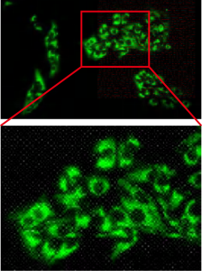
Olympus 20X objective
Mitochondria labeled with fluorescent dyes

Standard 10X objective
Stable mitochondrial cell line
5.Automatic imaging
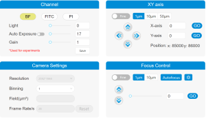
Equipped with a four-axis fully automatic motorized switching system, it can freely switch between bright field and dual fluorescence channels, with automatic focusing and automatic imaging according to the set program.
6.Intelligent algorithm
Intelligent anti-shake generates traceable videos.
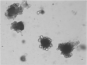
It supports the z-stack layer scanning of organoids and intelligent synthesis of clear images from different layers, scanning up to 40 layers at most.
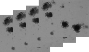
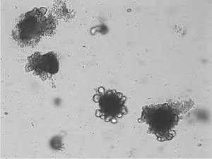
4X Olympus objective, Colorectal cancer organoid Z-stack synthesis
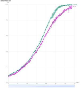
7.Data visualization
Matrix data overview, supporting data group analysis, automatic generation of proliferation curves, automatic display of cell confluence, scratch data analysis, transfection efficiency analysis, etc.
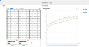
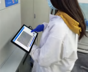
8.Three-terminal interconnection based on web architecture
The device is equipped with a high-performance host. Under the local area network, it supports remote settings and viewing of imaging data via mobile phones and tablets.


9.Support customization
It supports customized z-stack layer scanning, customized data analysis modes, etc.
Application
This instrument has a wide range of applications and features multi-channel fluorescence imaging, multi-site plate scanning, digital phase contrast, real-time imaging for generating cell growth curves, and other functions.
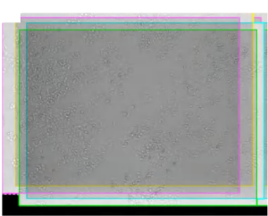
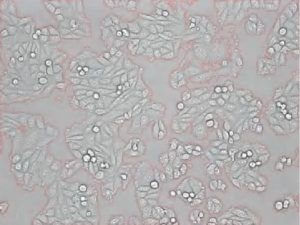
1.Bright field label-free live cell real-time imaging
It can be used for cell growth monitoring, cell culture quality control and optimization of culture conditions (such as medium development, etc.).
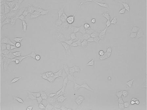
Bright field HEK293T cell imaging
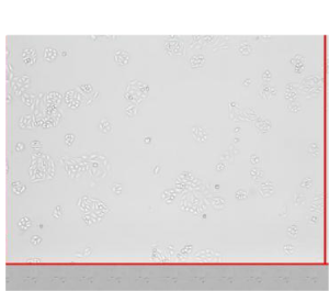
2.Cell proliferation and confluence analysis
CHO cell labeling
3.Scratch experiment imagingand analysis

4.Neuronal axon imaging
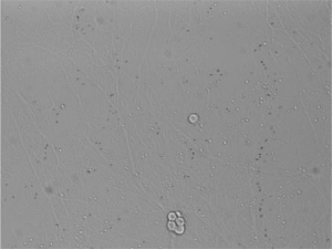
Bright field
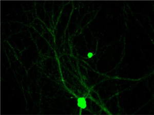
Imaging after fluorescence labeling
5.Analysis of transfection efficiency
![]()
![]()
![]()
![]()
![]()
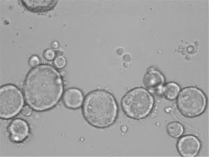
6.Organoid imaging
Specification
|
Item |
BCM3-300 |
BCM3-300F2 |
BCM3-300F3 |
|
Observation Method |
Bright Field |
2-channel fluorescence |
3-channel fluorescence |
|
Illumination |
LED, white |
LED, blue and green fluorescence light source |
LED, choose 3 of 4: blue, green, UV, red fluorescence light source |
|
Fluorescence Channel |
– |
Blue: EX 480/30nm, EM 535/40nm Green: EX 540/25nm, EM 620/60nm |
Choose 3 of 4: Blue: EX 480/30nm, EM 535/40nm Green: EX 540/25nm, EM 620/60nm UV: EX 375/40nm, EM: 450/40nm Red: EX630/15nm, EM: 680/20nm |
|
Objective |
Standard: 10X, NA 0.25. Optional: 4X/20X |
||
|
Field of View |
4X: 1.71mm*1.28mm; 10X: 0.68mm*0.51mm; 20X: 0.34mm*0.26mm |
||
|
Stage |
Motorized X/Y stage, 115mm*85mm, moving precision: 1μm, motorized Z-axis, precision: 1μm |
||
|
Adapted Container |
Compatible with 6-384-well plates and culture flasks of mainstream brands such as CORNING, etc. |
||
|
Camera |
High-sensitivity CMOS Camera, 5.0MP |
||
|
Imaging Mode |
Single field, 4-field, 9-field |
||
|
Power Supply |
100-240V, 1.4A, 50/60Hz 24V DC, 5.0A |
||
|
Computer |
CPU: i5; Memory: 16G DDR4; Hard disk: 4TB; Operating System: Windows 11; Monitor: 27inch, 4K |
||
|
Output Format |
Image: JPEG, TIFF, PNG; Video: AVI, MP4; Data: CSV |
||
|
Working Environment |
5~40°C, 20~95% RH, no condensation |
||
|
Dimensions |
328mm*242mm*350mm (W*D*H) |
||
|
Weight |
9.5kg |
10.5kg |
11.5kg |


