


BS-2084 Biological Microscope
Introduction
BS-2084 biological microscope is an innovative device that combines traditional microscopy observation with modern scanning technology. It not only retains the classic functions of a microscope but also serves as a digital scanning device for slide pathology scanning, greatly enhancing the working efficiency in the fields of research and medicine.
Details
Overview
Packaging & Delivery
Packaging Details:Strong Carton with Polyfoam Protection
Port:Beijing
Lead Time:Within 2-4 Weeks after Receiving Payment
Introduction
BS-2084 biological microscope is an innovative device that combines traditional microscopy observation with modern scanning technology. It not only retains the classic functions of a microscope but also serves as a digital scanning device for slide pathology scanning, greatly enhancing the working efficiency in the fields of research and medicine.
Features
1.High-quality real-time image acquisition
Equipped with a high-performance camera, the BS-2084 is capable of capturing high-definition images in real-time and supports photography function, allowing users to instantly save important observation results.
2.One-button fully automatic scanning imaging
One-button full-slide imaging, providing an overview of the entire slide information. Greatly improves work efficiency, helps reduce human errors, and enhances the accuracy and reliability of data.
3.Fully motorized XYZ axis manual-motorized integrated
Equipped with fully motorized X/Y/Z axis control, enabling precise and convenient sample positioning and focusing. Users can choose between manual adjustment or activate automatic scanning mode to adapt to different observation needs.
4.Compound illuminating system
Using a compound illuminating system to enhance contrast and effectively improve the uniformity of specimen illumination, ensuring uniform brightness across the entire field of view. Even at the edges of the field of view, uniform and bright background brightness can be achieved at any magnification.
5.LED light source, safe and environmental-friendly
Using LED light source, environmental-friendly and energy-efficient, providing soft and non-glaring illumination. Not only does it produce ideal imaging results, but it also reduces eye fatigue and enhances the observation experience.
6.Scanning Process
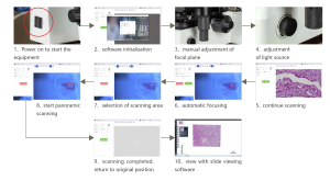
7.Concise exploration, convenient operating software
The operating software of the BS-2084 biological microscope achieves efficient microscopic observation with its intuitive user interface and advanced software control system. It ensures the accuracy and consistency of slide sample observation. Users can easily perform operations such as autofocus, automatic white balance, and automatic scanning, previewing real-time and capturing panoramic images automatically. The software also features flexible adjustment of exposure time and gain and supports single-layer or multi-layer image scanning, meeting various microscopic observation needs.
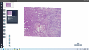
Multi-layer scanning stitching technology
By utilizing high-precision multi-layer scanning technology, it can achieve scanning of areas up to 50mm(X)*25mm(Y), automatically generating large area sample stitching images, providing users with a broader field of view.
Automatic focusing and white balance
To ensure image quality, the software features automatic focusing, which can quickly and accurately find the optimal focal plane. Meanwhile, the automatic white balance function ensures color accuracy and consistency, allowing accurate color information to be obtained under various lighting conditions.
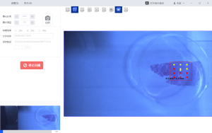
Automatic scanning and real-time preview
Users can observe real-time imaging of samples through the real-time preview function, facilitating the selection of regions of interest for automatic scanning, thereby saving time and improving scanning efficiency.
Exposure time and gain adjustment
Users can manually adjust the exposure time and gain according to the characteristics of the sample and imaging needs to obtain the best image quality.
Single-layer or multi-layer scanning
The software supports single-layer or multi-layer scanning, suitable for samples of different thicknesses and types, providing great flexibility and applicability.
Specification
|
Item |
Specification |
BS-2084 |
|
|
Optical System |
Infinite Color Corrected Optical System |
● |
|
|
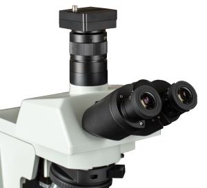
Viewing Head |
30° inclined infinity trinocular head, inverted image, interpupillary distance: 50-76mm, splitting ratio R:T=0:100 or 20:80 or 100:0 |
● |
|
|
Eyepiece |
High eyepoint wide field plan eyepiece PL10X/25mm, with adjustable diopter |
● |
|
|
Objective |
Infinity Plan Apochromatic Objectives |
4X/NA=0.16, WD=12.8mm |
○ |
|
10X/NA=0.4, WD=3.2mm |
○ |
||
|
20X/NA=0.75, WD=0.6mm |
● |
||
|
40X/NA=0.95, WD=0.15mm |
● |
||
|
60X/NA=0.9, WD=0.26mm |
○ |
||
|
(Oil) 100X/NA=1.35, WD=0.13mm |
○ |
||
|
Infinity Plan Super Apochromatic Objectives |
10X/NA=0.4, WD=3mm |
○ |
|
|
20X/NA=0.8, WD=0.6mm |
○ |
||
|
40X/NA=0.95, WD=0.18mm |
○ |
||
|
(Oil) 60X/NA=1.42, WD=0.17mm |
○ |
||
|
(Oil) 100X/NA=1.45, WD=0.14mm |
○ |
||
|
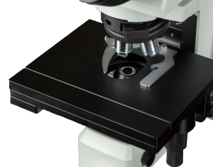
Nosepiece |
Inward quintuple bright field nosepiece, with DIC slot |
● |
|
|
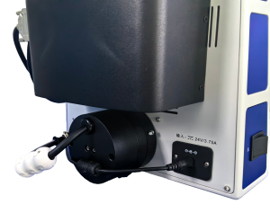
Microscope Body |
Compound eye lighting system, manual and automatic integrated biological frame, including low position coaxial focusing mechanism and automatic focusing mechanism, coarse range: 15mm, fine precision: 0.001mm, automatic focusing range: 3mm, repeated positioning accuracy <1um. Equipped with upper limit and tension adjustment. Brightness continuously adjustable. Including 1.3 mega-pixel macro camera for positioning in the scanning area. |
● |
|
|
Wide voltage adapter, grounded, input 100V-240V, output 24V/3.75A |
○ |
||
|
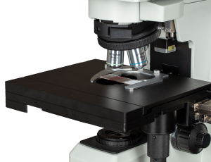
Stage |
Manual and automatic integrated three-layer composite electric stage, stage size: 260mm(X)*221mm(Y), moving range:100mm(X)*50mm(Y), positioning accuracy<5um, repetitive positioning accuracy<±3um. Maximum speed:20mm/s |
● |
|
|
Condenser |
Long working distance abbe condenser, NA >=0.65, with variable aperture diaphragm |
● |
|
|
Transmitted Illumination |
Wide band 3W LED lamp house (cool color, color temperature 4700-5500K) |
● |
|
|
Camera |
7 mega-pixel high-speed scanning camera, color camera, target size: 1.1” (14.4mm*9.9mm), frame rate: 51.4fps, resolution ratio: 3200*2200, USB 3.0, pixel size: 4.5um*4.5um |
● |
|
|
Software |
Scanning software: Real-time image acquisition and photography. Automatic white balance, brightness adjustment and gain adjustment. Measuring, measure scaling and dimension, measurement data can be exported. PC software control of microscope electric function (X/Y/Z axis). Autofocus, stitching of up to 50mm(X)*25mm(Y) area. The software include Dongle. |
● |
|
|
PC |
DELL Rrecision T3660. CPU: Intel i7 12700. Memory: 32GB. Graphics card: RTX1660S. Display: P2722H (Resolution rate 1920*1080). Hard disk: 1TB SATA, SSD 256 GB. Mainboard: three USB3.0 interfaces. Operating system: Windows 10X64 |
● |
|
|
Adapter |
1X C-mount adapter, focus adjustable |
○ |
|
Note: ● Standard Outfit, ○ Optional
Accessories
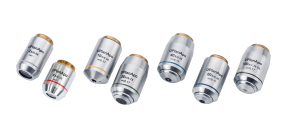
1.Infinity Plan Apochromatic Objective
Infinity corrected plan apochromatic objectives correct axial chromatic aberrations in red, green, and blue light, causing them to converge on the same focal plane. They effectively correct spherical aberration in purple light, faithfully reproducing the colors of the observed objects, such as red, green, and blue. With an extra-large numerical aperture, they further enhance resolution and effective magnification.
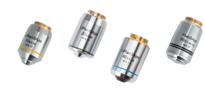
2.Infinity Plan Super Apochromatic Objective
Infinity corrected plan super apochromatic objectives adopt innovative design concepts and break through the technological bottleneck of unconventional-shaped lens elements. They maximize the capture of edge light within a 45mm coaxial zone, resulting in more uniform, brighter, and higher-resolution fluorescence images under equivalent conditions.
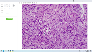
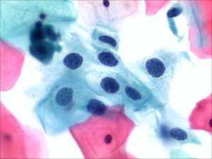
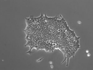
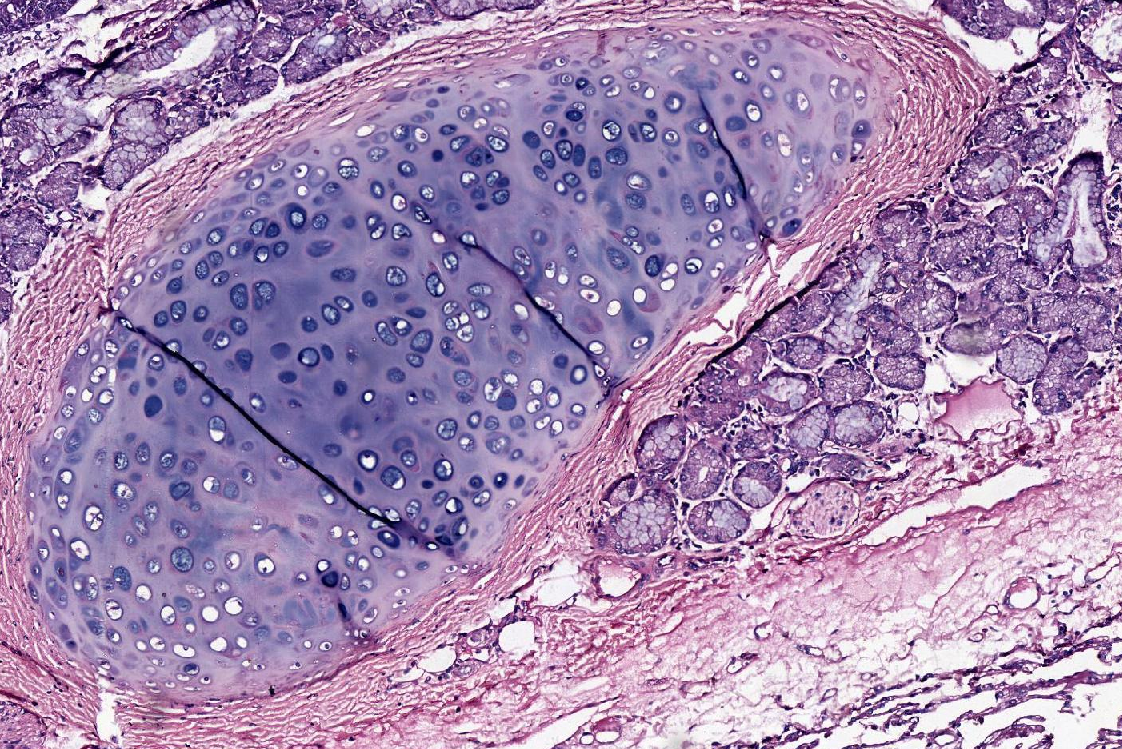
Sample Image


")
")


