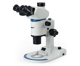

Dissecting Microscope – Definition, Principle, Parts, Uses
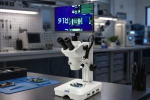 The dissecting microscope, also known as the stereo microscope, provides you highly three-dimensional upright images through a dual-path optical system to show the depth and details of samples. The dissecting microscopes are widely used in dissection of small animals and tissue samples in biological laboratory experiments and teaching, as well as in industrial inspections to check circuit boards and welding. Let’s delve deeper into the dissecting microscope and get some suggestions for choosing one.
Definition – What is Dissecting Microscope
The dissecting microscope is an optical microscope with a low magnification, typically between 5X to 250X. The dissecting microscope is also known as a stereo microscope. It provides you 3D upright images through a dual-path system, allowing you to perceive the depth of the sample. It often used for dissection.
Principle – How to Get 3D Effect Image
The dissecting microscope consists of eyepieces, internal lenses and objectives to form a complete optical system. After the sample is magnified for the first time by the objective, the image is reversed into upright by the prism under the eyepieces, and then magnified again by the eyepieces. Finally, you can get the upright image of the sample in the eyepieces. Simple dissecting microscopes only provide a fixed magnification, while advanced dissecting microscopes can be zoomed. Zooming is achieved by the intermediate lens group between the objective and the eyepieces in the microscope body. The objective is responsible for the main magnification effect. Rotating the zoom knob on the side of the microscope body can move the position of the intermediate lens group and the magnification change is achieved by adjusting the distance between the intermediate lenses.
There are two optical path designs for dissecting microscopes, Greenough Optical System and Galilean Parallel Optical System. The core mechanism of imaging of the dissecting microscope is based on the dual optical path system, observing the sample from different angles through two independent optical paths to simulate the parallax of human eyes. The left eyepiece and the right eyepiece respectively form the images and then human brain fuses these two images to form a 3D stereoscopic image with depth.
The Greenough optical system uses two complete symmetrically tilted optical systems. There are two completely independent optical paths in a V shape with an angle of 12°-15°. So, the microscope body above the objective can be designed as a slender shape and the microscope body is smooth in lines and small, convenient for assembly and use. The design of two tilted optical systems can simultaneously achieve high flatness and deep focus depth, correctly focusing samples with large concavities and convexities and present perfect stereoscopic images. With the Greenough optical system, the dissecting microscope can ensure the basic stereoscopic imaging while significantly reduces the size to save space and is lightweight and easy to carry, suitable for outdoor uses and experimental benches with limited space.
The Galilean parallel optical system, also known as CMO optical system, is a parallel optical system, using one large Common Main Objective in front, followed by two optical paths with no difference and parallel to each other, significantly improving image quality. At the same time, other accessories such as fluorescence attachment and coaxial illuminator can be inserted in the parallel optical paths without affecting imaging. The Galilean parallel optical system has better color reproduction capability, higher image resolution and smaller aberration. It is suitable for applications required high-performance imaging and you can flexibly add accessories to achieve different imaging effects as needed.
Parts – What Constitutes a Dissecting Microscope
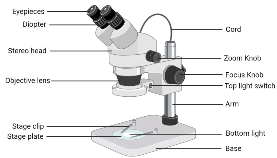
The main components of a dissecting microscope include the microscope head, the base and stand, and the focusing holder. Among them, the microscope head contains the most critical optical components, including the eyepieces, the objective and the internal prism and lenses, etc.
Head – Objective
The main lens in the optical system. The objective collects light from the sample and forms the first magnified image.
Head – Eyepiece
The lenses used to magnify and observe the formed image. Common eyepieces include 10X and 15X.
Head – Internal Prism & Lenses
The prism below the eyepiece reverses the image to make the image observed in the eyepiece upright. In the zoom dissecting microscope, there is a continuously variable zoom lens group that changes the magnification by altering the distance between the intermediate lenses. A trinocular viewing head can be connected to a microscope camera through C-mount interface to capture or share images.
Base & stand
The base is the position for placing the sample. A large base can accommodate large samples and a base with an LED light source can provide a transmitted illumination. The column supports the microscope head, connecting the base and the focusing holder.
Focusing holder
The focusing holder is used to move the head up and down, helping the objective to focus the sample to get a clear image. Some dissecting microscopes have a focusing system that moves the stage up and down to make the objective focus the sample.
Illuminator
The dissecting microscopes generally use LED light sources. The transmitted illumination is usually on the base, and the incident illumination usually on the stand or on the focusing holder. Transmitted light passes through the sample from below and then directed upward to enter the objective, generally used for transparent or semi-transparent samples, such as to plant sections, small transparent invertebrates, slides, etc. For non-transparent samples, the transmitted light only shows the outline of the sample and can be used for checking the outline of workpieces. Incident light is directed downward onto the sample and then reflect back, entering the objective, generally used for observing non-transparent samples, such as insects, PCBs, rock sections, etc. Incident illumination is also used in dissection. You can choose other light sources according to your needs, such as LED ring lights, fiber optic gooseneck light, etc.
Uses – Why and Where Do You Use a Dissecting Microscope
The advantages of the dissecting microscopes include:
3D Images
The dissecting microscope can provide a 3D image of the sample with a large depth of field, which is helpful for clear observation of depth, height and surface structure.
Long Working Distance
Although the magnification of the dissecting microscope is usually relatively low, it has a relatively long working distance. The distance between the objective and the sample is large, providing sufficient space for convenient operation, such as using tweezers, needles, scalpels or other tools for operation. You can dissect plants or small animals, weld, set gemstones or do other things under the dissecting microscope.
Upright Image
The dissecting microscope reverses the image through the prism below the eyepieces to form an upright image. The image seen in the eyepiece is in the same direction as the actual sample, which is convenient for the operations under the microscope and avoid confusion of movement directions.
Simple Operation
The dissecting microscope has a simple structure and is easy to operate. There are small models that is convenient to carry outdoor for use.
Rich Accessories Available
According to different needs, there are a wide range of illuminators, stands and attachments available for selection, suitable for different types of samples and different observation methods.
The applications of the dissecting microscopes are very extensive.
Teaching
It is used for students to study animal and plant specimens, dissect animals and plants, observe the germination process, etc. It has a high cost-performance ratio and is easy to operate.
Biology
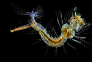
It can be used for animal and plant dissection, insect research, embryo observation, etc.
Crime Investigation
It is used to observe evidences such as hair, fibers, soil, bullet fragments, fingerprints, marks, etc. It can also be used for document inspection.
Industrial Manufacturing
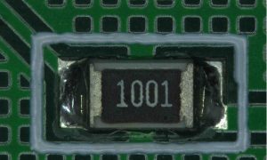
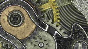
It is used in industrial manufacturing and quality control, such as circuit boards, electronic components, watches, small mechanical parts, etc.
Gem & Collection
It is used for gem research and grading. With dissecting microscope, you can see tiny impurities inside the gem and surface defects. It can also be used for checking the surface marks, luster and texture of coins, stamps and other collections.
Paleobiology & Geology
It is used for cleaning and inspecting fossils, and can also be used for observing rocks, minerals and crystals.
Textile Industry
It is used to inspect the quality of fibers, fabric and textile materials.

Products – Which of the Dissecting Microscopes is More Suitable?
Choose a suitable dissecting microscope depend on your needs.
The dissecting microscope with Greenough optical system is designed compactly with a small size, making it easy to carry. The basic models are suitable for teaching and other occasions that require high cost-effectiveness.
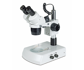
BS-3016A1 Binocular Stereo Microscope
The dissecting microscope can provide stereoscopic images. With a long working distance, it is suitable for operations under microscopes.
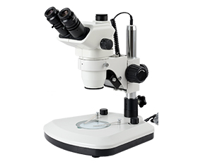
BS-3044A Binocular Zoom Stereo Microscope
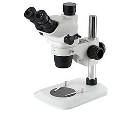
BS-3047 Zoom Stereo Microscope
BS-3049B Binocular Zoom Stereo Microscope
The dissecting microscope with Galilean parallel optical system can provide high-resolution, good color reproduction and low distortion imaging, suitable for those with high requirements for image quality.
BS-3090 Parallel Light Zoom Stereo Microscope
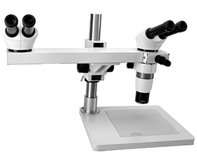
The dissecting microscope with Galilean parallel optical system can also be inserted accessories as needed, without affecting the imaging quality. It can meet different observation methods such as fluorescence, dark field, simple polarizing, multi-view observation, etc.
BS-3060C Binocular Zoom Stereo Microscope
We have a wide range of accessories for the dissecting microscope to meet your different need, including illuminators, stands, stages, etc.