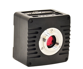

BestScope Pathology Slide Scanner Optimizes Your Workflow
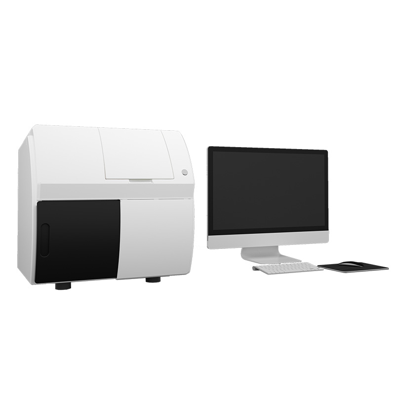
A pathological slide scanner is composed of a slide loading system, microscopic optical system, control system, scanning camera, precise scanning platform and application software. It effectively overcomes the limitations of traditional microscopes in terms of time, labor and geographic constraints
With simple slide loading, the whole slide scanner system will automatically complete the slide loading, label recognition, tissue region detection, automatic focusing, automatic scanning, and finally present the pathological slides in digital images on the computer monitor. You can view the entire slide, zoom in on regions of interest , annotation, measurement on the digital slides, and support for individual saving and transmission.
This solution meets various demands, including hospitals, clinics, large-scale research laboratories, teaching institutions and remote pathology diagnosis.
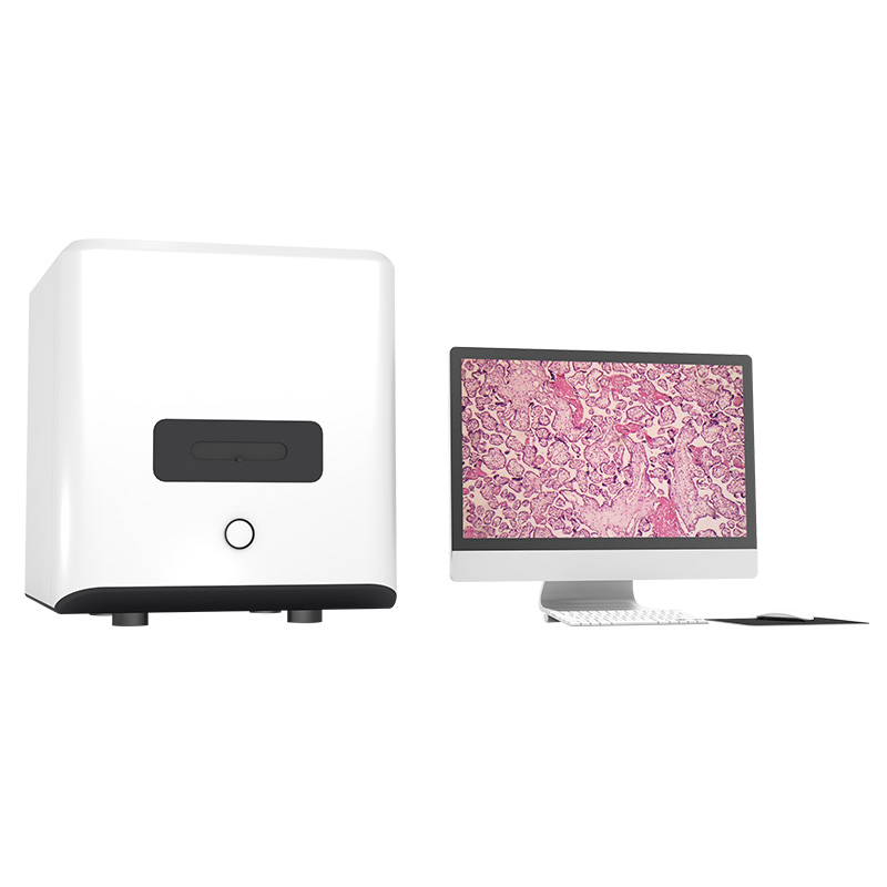
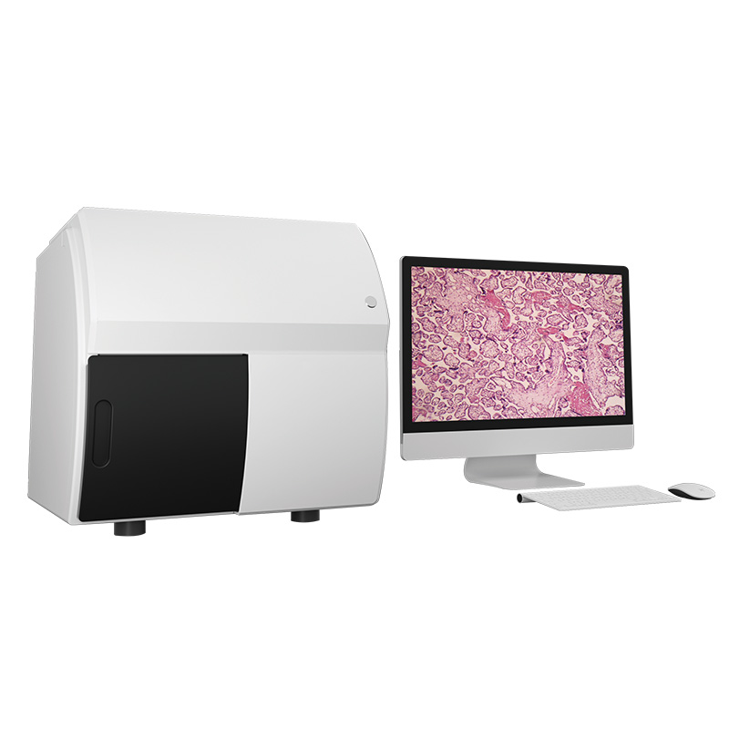
Scanpro2 Series Pathology Section Scanner
Scanpro2 series pathology section scanner employs advanced scanning technology, enabling it to complete the efficient scanning of 15mm*15mm slides in just 20 seconds, significantly enhancing the speed and accuracy of pathological diagnosis. Furthermore, it is capable of precisely scanning all types of pathological slides—whether they are lightly stained slides, uniquely shaped "island" slides, or specialized samples such as Tissue Microarrays (TMAs), standard tissue slides, and large-format tissue slides (2-inch or 4-inch). Concurrently, Scanpro2 delivers high-quality scans; its high-fidelity color reproduction technology ensures the accurate rendering of colors when pathologists, histologists, biologists, hematologists, veterinary specialists, or other medical professionals examine the slides.
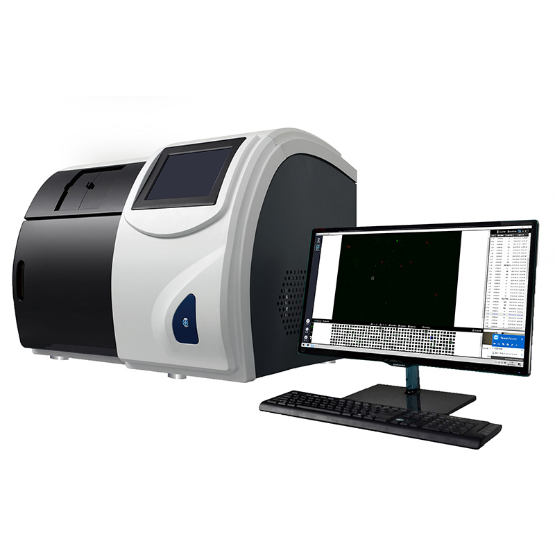
Scanpro3 Series Mycobacterium Tuberculosis Microscopy Scanning System
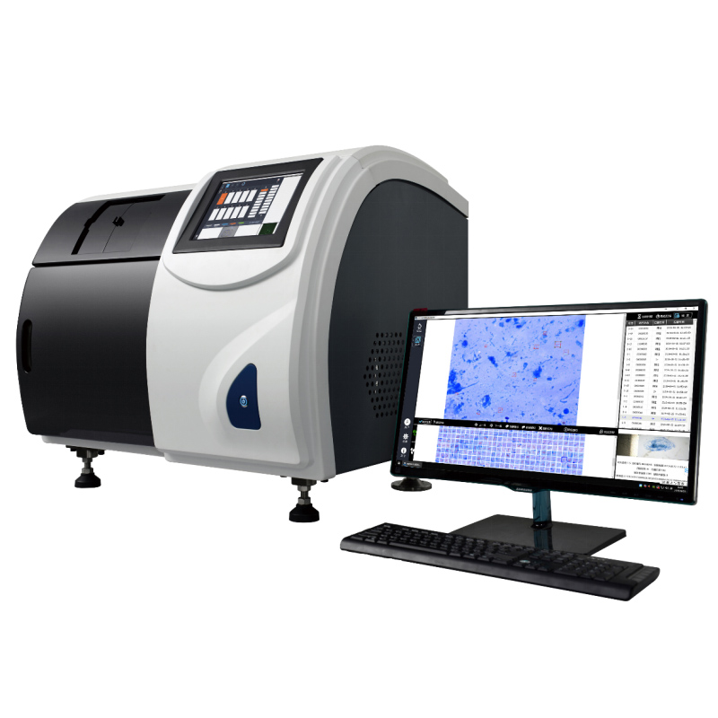
Scanpro3 Series is an intelligent and professional screening system designed for the detection of Mycobacterium tuberculosis (TB). It integrates high-resolution digital microscopy with a high-precision motion control system. The system performs automated scanning, image acquisition, and intelligent analysis of smear samples. Scanpro3 Series significantly enhances operational efficiency in tuberculosis screening and medical laboratory workflows; particularly in negative/positive grading. Pathologists only need to perform a secondary manual review of suspected bacilli to complete the diagnostic process.
Quality Assurance

ISO9001, ISO13485, ISO14001 Management System Certificate.

Most of the products are CE certified.

Big quantity orders can be delivered in a short time.

All the products are provided with 3 years warranty.
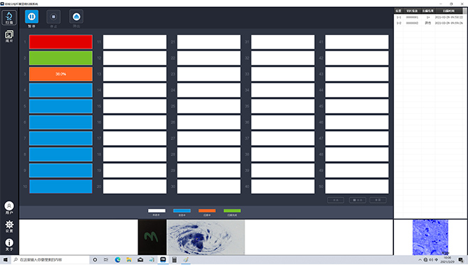
Scanpro3 Series Automated TB Microscopy Step-by-Step Demo
Automatic image collection and storage, intelligent identificationanalysis and processing of sputum smears
The Scanpro5 series slide scanning system adopts patented area scanning technology. It can scan slides with just one-click and the operation is simple. Through precise hardware control, stable operation is achieved. Through software and intelligent platform, system expansion can be realized, creating an intelligent diagnostic system and digital platform.
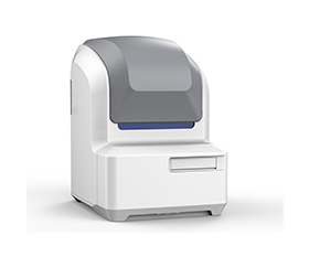
The Scanpro5 series slide scanning system adopts patented area scanning technology. It can scan slides with just one-click and the operation is simple. Through precise hardware control, stable operation is achieved. Through software and intelligent platform, system expansion can be realized, creating an intelligent diagnostic system and digital platform.
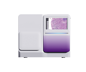
The Scanpro5 series slide scanning system adopts patented area scanning technology. It can scan slides with just one-click and the operation is simple. Through precise hardware control, stable operation is achieved. Through software and intelligent platform, system expansion can be realized, creating an intelligent diagnostic system and digital platform.
- Scanpro4
- Application
- Specification
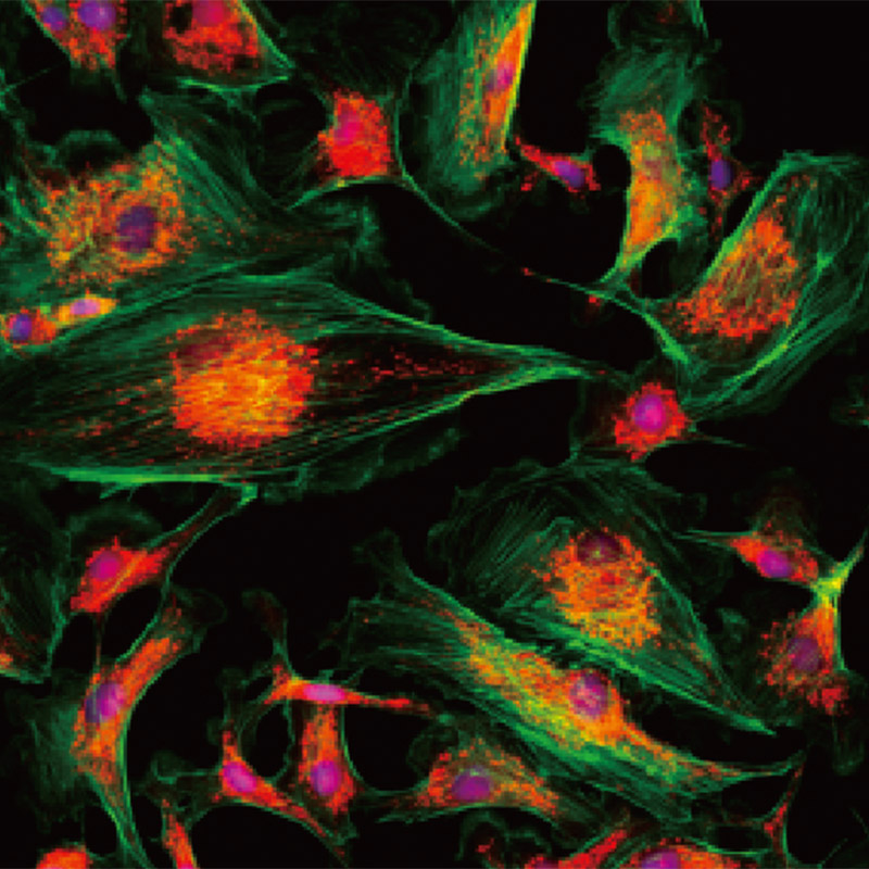
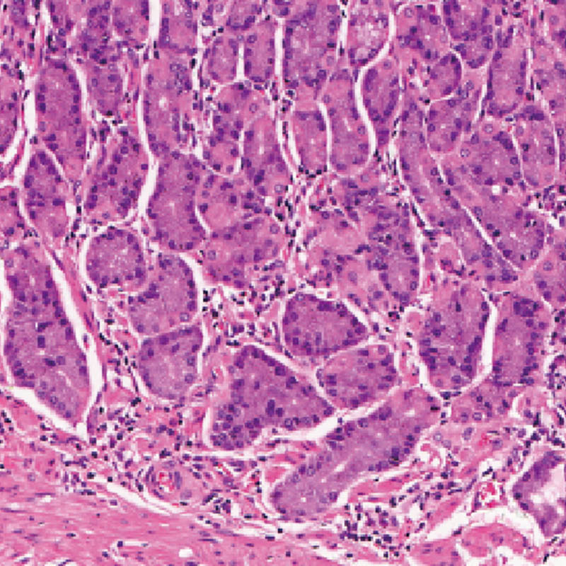
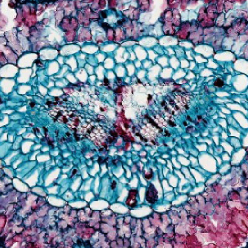
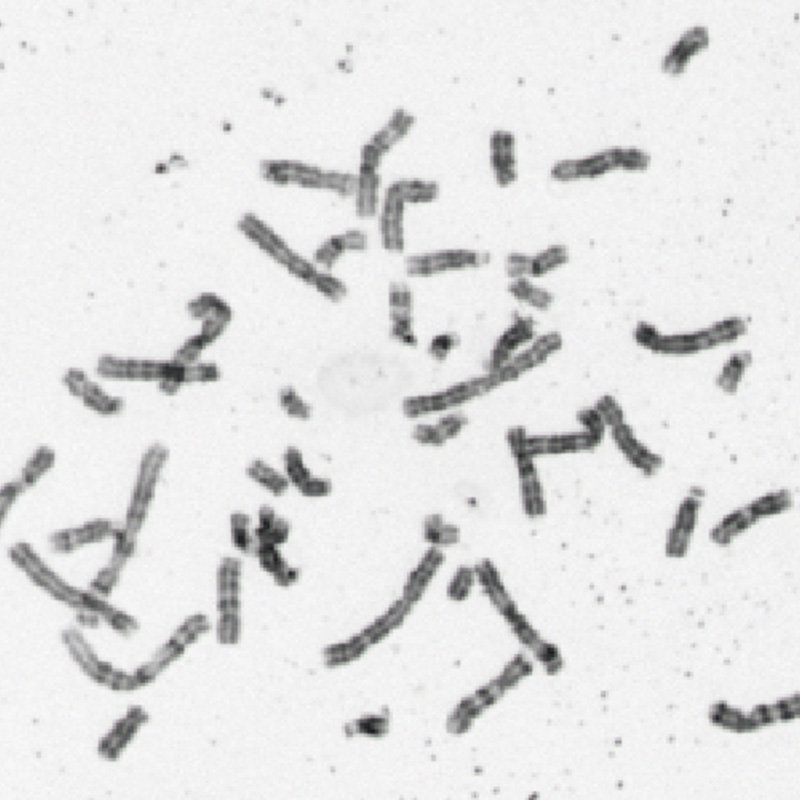
Microorganism
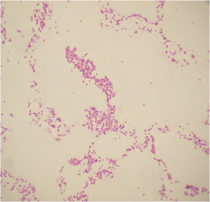
Gram-negative bacteria Escherichia coli
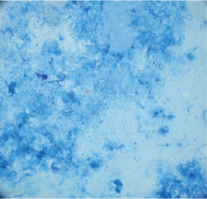
Nissl staining
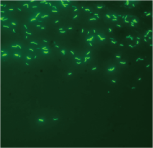
Auramine O staining
Pathology
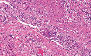
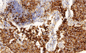
FISH fluorescence
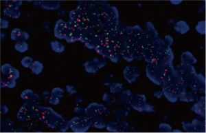
Multicolor fluorescence fusion image
Blood
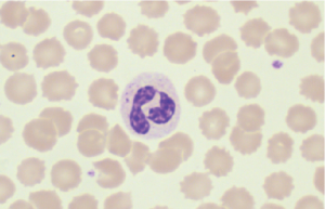
Blood cell morphology analysis
|
Item |
Specification |
|
Observable Sample |
Slides with/without standard coverslips |
|
Slide Size |
Standard slide trays (L*W*H, 5 slides): 75mm-76.5mm (2.95in-3in), 25mm-26.5mm (0.98in-1in), 0.9mm-1.2mm (0.04in inches-0.05 inches) |
|
Optional slide tray 1 (L*W*H, 2 slides): 75mm-76.5mm (2.95in-3in), 51mm to 53mm (2 in-2.09in), 0.9mm-1.2mm (0.04 inches-0.05 inches) |
|
|
Optional slide tray 2 (L*W*H, 1 slide): 100mm-102mm (3.94in-4.02in), 75mm-76.5mm (2.95in-3in), 0.9-1.2mm (0.04 inches-0.05 inches) |
|
|
Observation Method |
Bright field |
|
Fluorescence |
|
|
Illuminator |
Built-in transmitted light Kohler illumination, high intensity and high color rendering LED (up to 50,000 hours lifetime) |
|
Objective Lens |
Compatible objectives: 10X, 20X, 40X, 60X and 100X. 3-position motorized turret (includes select oil immersion) |
|
Automatic oil distributor |
|
|
Stage |
Automatically controlled motorized XY stage |
|
Focusing |
Automatically controlled autofocus with support for focused topographic maps and real-time autofocus |
|
Scan Camera |
Color camera, 1.1” CMOS, 4.5um*4.5um pixels, 7 mega-pixels, high sensitivity, high resolution |
|
Monochrome camera, 1.1” CMOS, 4.5um*4.5um pixels, 7 mega-pixels, high sensitivity, high resolution |
|
|
Panoramic Camera |
Color Camera, 5 mega-pixels |
|
Capacity |
5 slides: 1 slide tray, up to 5 slides, can be upgraded to multi-tray feeding mechanism |
|
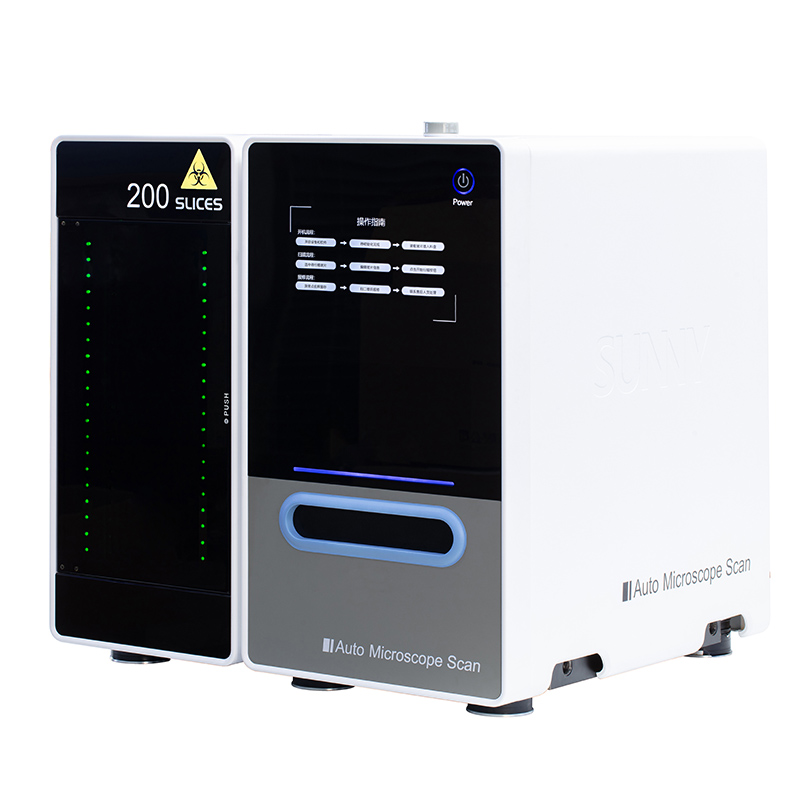
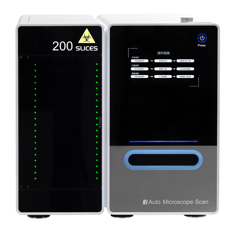
100/200 slides: up to 40 slides trays, up to 200 slides |
|
|
400/600 pieces can be customized |
|
|
Pixel Resolution |
10X (NA 0.4): 0.672 um/pixel |
|
20X (NA 0.8): 0.335um/pixel |
|
|
40X (NA 0.95): 0.168um/pixel |
|
|
60X (NA 1.4): 0.112um/pixel |
|
|
100X (NA 1.35): 0.067um/pixel |
|
|
Scanning Time |
Bright field is less than 60 seconds (20X objective lens, scanning area 15mm*15mm) |
|
Software |
Auto barcode reading, auto focus topographic map, auto scan, auto stitching, pause and resume scanning, Z-stack imaging, multiple image formats (BMP, JPEG and TIFF), simultaneous multi-image display, stepless zoom, upload while scanning slide browsing, annotation, screen capture |
|
Fluorescence |
Equipped with professional LED fluorescent illuminator, motorized 4-hole fluorescent reflected lens turntable, motorized filter wheel, fluorescent light source |
|
Oil component |
Long life, maintenance-free, 5 million times |
|
Weight |
Slide scanner of 5 slides: 40kg, slide scanner of 200 slides: 70kg. 1 slide tray: 0.1 kg |
|
Operating Environment |
Temperature: 10°C-30°C. Humidity: up to 80% (non-condensing) |
|
Power Consumption |
220W |
|
Power Rating |
Input: 220V AC, 50Hz, 1A |
Related Product