Different Types of Optical Microscopes
Microscopes are widely utilized across various fields. To meet the diverse needs of these fields, microscopes are classified into several types. Among them, electron microscopes and optical microscopes are two important classifications. Electron microscopes use electron beams as the light source and they are primarily used in the study of molecular structures in life sciences, analysis of crystal structures in materials science, and applications in nanotechnology in physics. On the other hand, optical microscopes use visible light as the light source. This article will provide detailed classification based on the different characteristics of microscopes.
Classification by Microscope Application
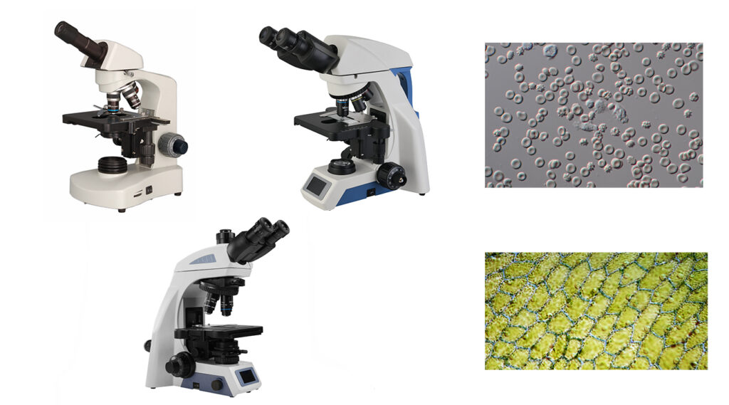
Biological Microscope/Compound Microscope
The biological microscope is the most common type of compound microscope. It utilizes objective and eyepiece lenses to magnify specimens, enabling the naked eye to observe tiny objects. The maximum total magnification can reach up to 1600X. Its resolution is determined by the numerical aperture(NA) and wavelength, where a larger numerical aperture(NA) allows more light to enter, resulting in higher resolution. Conversely, shorter wavelengths yield higher resolution due to their smaller diffraction limits.
Resolution=0.61λ/NA
The biological microscope is used for observing microorganisms, cellular structures and functions, bacteria, tissue cultures, pathological analysis, and finds widespread applications in education, healthcare, laboratory research, and other fields.
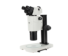
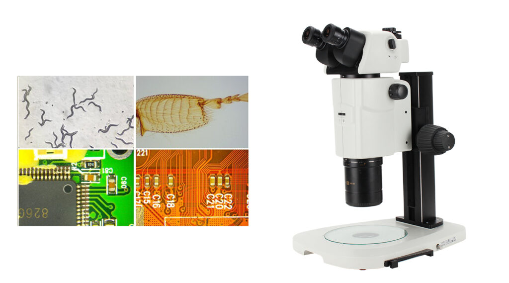
Stereo Microscope/Dissecting Microscope
The stereo microscope, also known as a dissecting microscope, is so named because it provides a three-dimensional view of specimens. Unlike the compound microscope with its transmitted illumination, the stereo microscope uses reflected illumination, and each eyepiece corresponds to an independent optical path. This allows for the observation of specimens in 3D. The magnification range of stereo microscopes is typically lower, usually between 5x and 50x. They are commonly used for circuit board repair, electronic component analysis, jewelry restoration, and the dissection of insects and flowers. The working distance is an important parameter for stereo microscopes, as a longer working distance facilitates precise operation under the microscope.
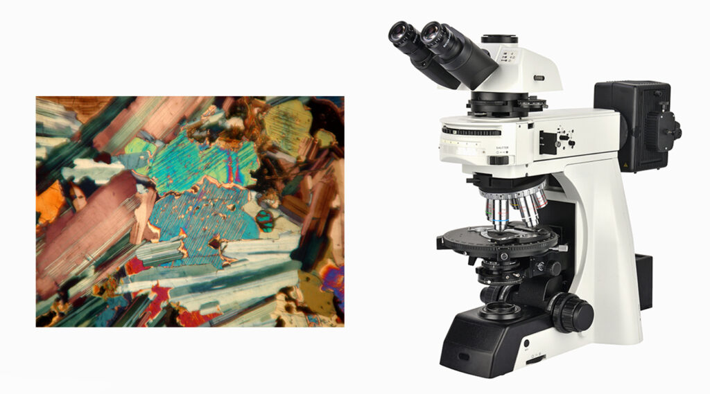
Polarizing Microscope
The polarizing microscope utilizes the principle of polarization to observe materials exhibiting birefringence(double reflection), either through transmission or reflection. Birefringence is a fundamental characteristic of crystals. Therefore, polarizing microscopes find widespread applications in fields such as minerals, rocks, polymers, fibers, glass, semiconductors, and chemistry. It can be further divided into petrographic microscope and ore microscope, with the difference being that the ore microscope requires a reflected illumination.
The basic principle of a polarizing microscope involves polarization devices – a polarizer and an analyzer. The polarizer, positioned in the optical path between the light source and the specimen stage, converts natural light into polarized light with a single vibrational direction. The analyzer, located between the objective and eyepiece, is used to analyze the specimen’s response to polarized light, detecting phenomena such as birefringence. This enables the observation and analysis of the specimen’s characteristics.
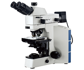
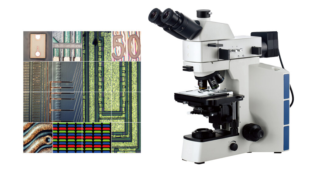
Metallurgical Microscope
The metallographic microscope, also known as a metallurgical microscope, is a type of compound microscope that shares similarities with the biological microscope in appearance but differs in several aspects. Primarily used for observing opaque materials such as metals, alloys, ceramics, and polymers, the metallographic microscope typically employs a reflected light source. This illuminates the specimen surface effectively from above, enhancing contrast for observation and analysis.
While the metallographic microscope is mainly utilized for observing opaque materials, some specimens require transmitted light to examine their internal structures. Therefore, certain microscopes are equipped with both reflected and transmitted illumination to cater to different needs.
Objective lenses typically come in magnifications such as 5X, 10X, 50X, and 100X, with a long working distance designed specifically for observing opaque specimens.
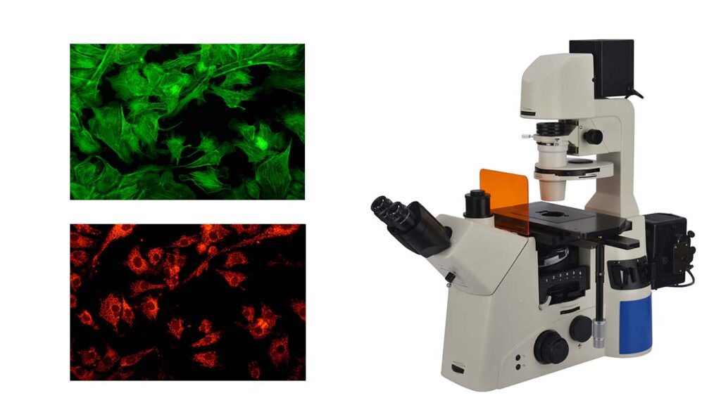
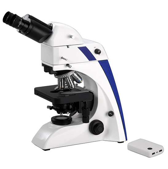
Fluorescence Microscopy
Fluorescence microscopy utilizes short-wavelength light (excitation light source such as ultraviolet or blue light) to illuminate specimens labeled with fluorescent dyes, inducing them to emit longer-wavelength fluorescence upon excitation. It is primarily used in live cell research, protein characterization, antigen identification, and immunoprotein identification.
The key components of a fluorescence microscope include the excitation light source, filter sets, and beam splitter. Mercury lamps, xenon lamps, and LEDs are commonly used as the main light sources. Mercury lamps offer high intensity and broad spectral coverage suitable for exciting various fluorescent substances, but they have long preheating times and limited lifespans. Xenon lamps provide stable output and longer lifespans compared to mercury lamps. LEDs offer stable illumination, generate less heat, have long lifespans, and are energy-efficient, making them increasingly popular.
Filter sets consist of excitation and emission filters. Excitation filters are used to select filters that transmit excitation light, blocking non-specific wavelengths and allowing only specific wavelengths to pass through, ensuring proper excitation of fluorescent molecules. Emission filters only allow wavelengths emitted by fluorescent dyes to pass through, blocking other unwanted light, especially excitation light, thereby enhancing imaging contrast and clarity.
A beam splitter is a special optical lens that separates excitation light from emitted light, reflecting excitation light to fluorescent dyes and transmitting the emitted light to the emission filter.
Epifluorescence microscopy, a common type of fluorescence microscopy, projects light vertically onto the specimen surface, utilizing reflection and refraction principles to magnify images, making it more suitable for observing opaque and semi-transparent specimens.
Confocal microscopy is a special type of fluorescence microscopy that uses lasers as light sources. The light beam is focused through a lens onto a very small spot on the specimen surface. The spot is scanned horizontally and vertically on the specimen through a scanning system to achieve three-dimensional imaging. When the laser spot illuminates the fluorescent dye, the fluorescence signal is captured by a detector, forming a fluorescence image.
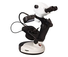
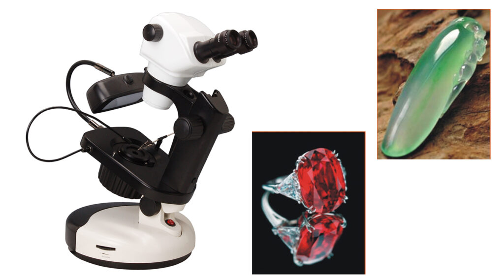
Gemological Microscope
The gemological microscope is a type of stereoscopic zoom microscope commonly used for magnifications ranging from 10X to 80X. It is equipped with both bottom and top illumination sources to ensure uniform lighting of the specimen. Gemstone holders are placed on both sides of the stage to stabilize the specimen. This microscope is specifically designed for observing and identifying gemstones and jewelry.
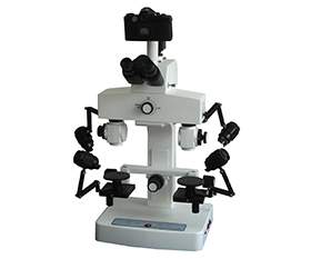
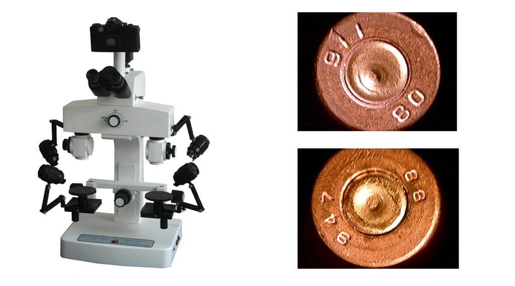
Comparison Microscope
The comparison microscope is a type of microscope used for comparative analysis of similar items. It features two separate optical paths, each with its own objective lens, eyepiece, and illumination. This setup allows for simultaneous observation of two specimens placed side by side. Comparison microscopes are commonly used in forensic science, ballistic analysis, art authentication and restoration, document examination, and other fields requiring detailed comparisons between specimens.
Classification by Microscope Observation Method
Brightfield Microscopy
Brightfield microscopy is the most common type of microscope, used for observing specimens against a bright background. It is primarily employed for observing transparent specimens such as cells, bacteria, microorganisms, pathological tissues, blood specimens, and similar materials.
Darkfield Microscopy
Darkfield microscopy operates on a different principle than brightfield microscopy. Darkfield microscopy utilizes off-axis or oblique illumination techniques, often employing a darkfield condenser. This condenser prevents direct light from entering the objective lens, allowing only light scattered or diffracted by the specimen to enter the objective lens at an oblique angle. This illumination technique effectively darkens the background surrounding the specimen while making the specimen appear bright, thereby enhancing image contrast. Darkfield microscopy is particularly useful for observing surface features of transparent specimens and unstained specimens, such as live microorganisms, viruses, and cellular structures.

Phase Contrast Microscopy
Phase contrast microscopy utilizes the interference phenomenon of light to make phase differences in transparent specimens visible. The human eye can only perceive differences in wavelength (color) and amplitude (brightness) of light. For transparent specimens, as light passes through, there is minimal change in wavelength and amplitude, making small phase differences imperceptible to the eye. However, by using a phase plate and phase-contrast objectives, phase contrast microscopy converts the phase differences generated by the specimen into differences in brightness that are perceptible to the eye. This enhances the visibility of specimen structures, making it suitable for observing live cells and unstained tissue sections.
DIC Microscope (Differential Interference Contrast Microscope)
The Differential Interference Contrast Microscope (DIC Microscope) is not only suitable for observing colorless transparent objects but also presents images with a relief-like three-dimensional appearance, offering a more realistic view compared to phase contrast microscopy. DIC microscopy utilizes polarized light and includes four special optical components: a polarizer, DIC prism, DIC slider, and analyzer. Initially, light passes through the polarizer, becoming polarized light. Then, the light is split into two beams by the prism. These two beams undergo different phase shifts upon interaction with the specimen. After passing through the specimen the light beams are recombined by the objective lens, generating interference based on the phase difference. As a result, the light distribution at different heights of the specimen exhibits high-contrast brightness differences, reflecting the relief-like three-dimensional appearance of the specimen in the image.
The different observation methods of polarizing microscopy and fluorescence microscopy were mentioned in the previous classification of microscope application fields, and they are not described in detail here.
Classification by Microscope Structure
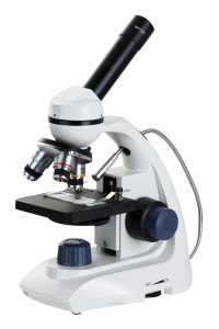
Monocular Microscope
The monocular microscope is a traditional microscope that allows observation of specimens through a single eyepiece. Despite having only one eyepiece, monocular microscopes offer clear and detailed views of biological specimens and materials. Its advantages lie in its simple structure, portability, and affordability, it is suitable for basic education and preliminary research.
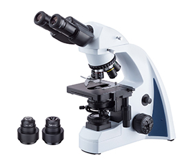
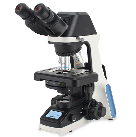
Binocular Microscope
The binocular microscope is a microscope with two eyepieces. Both eyes can observe the specimen at the same time, which is closer to the way we usually see things and can reduce visual fatigue. Most types of microscopes on the market are binocular microscopes, which are suitable for higher education, medical diagnosis, professional laboratories, scientific research and other fields.
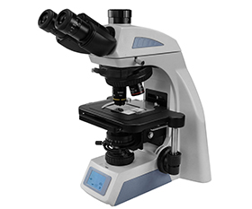
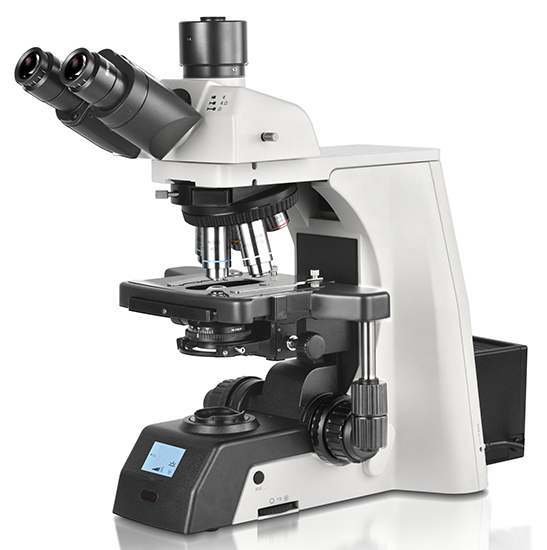
Trinocular Microscope
The trinocular microscope is an extension of a binocular microscope, featuring an additional third eyepiece tube. However, this third eyepiece tube is not used for direct observation of the specimen, instead, it is connected to a microscope camera to capture images and videos of the specimen. These images and videos can then be analyzed and processed using software. The optical pathway for this part typically includes a beam splitter for switching, allowing observation through both eyepieces and the device connected to the third eyepiece tube. Trinocular microscopes are particularly useful for situations where image documentation and analysis are required.
Upright Microscope
The upright microscope is a common type of optical microscope, where the objective lens is positioned above the specimen, and the light source is located at the base of the microscope. Light passes through the specimen from below via the condenser and is observed through the eyepiece. Due to the relatively small working distance between the objective lens and the specimen, upright microscopes are suitable for observing thin objects.
Upright microscopes can be configured with different observation modes according to requirements, such as brightfield, darkfield, phase contrast, and fluorescence microscopy.
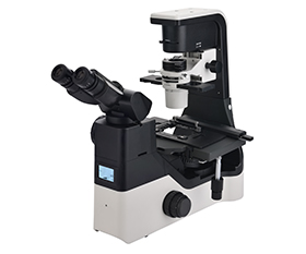
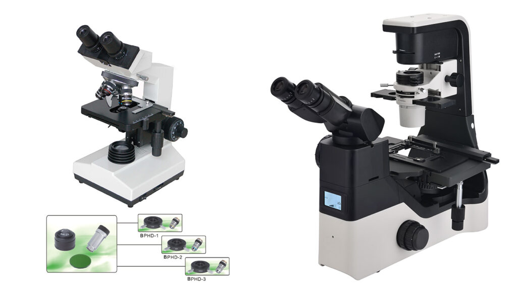
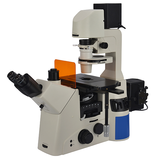
Inverted Microscope
The inverted microscope can be divided into biological inverted microscope and metallurgical inverted microscope. Their main feature is that the objective lens is positioned below the specimen stage, while the illumination system and condenser are located above the specimen stage. This design is suitable for observing thick and large specimens and facilitates operation, such as observing live specimens in culture dishes and wafer inspection.
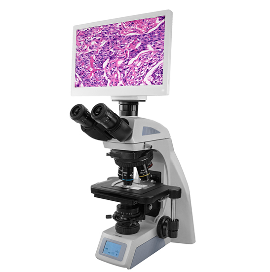
Digital Microscope
The digital microscope utilizes built-in or external microscope cameras to convert optical signals into digital signals, allowing images to be directly observed on a computer or HDMI monitor. They enable real-time image capture, storage, and sharing and they can also perform editing, analysis, and measurement functions through software. Digital microscopes are widely used in various fields.