What is Laser Scanning Confocal Microscopy?
Confocal microscopy is a technique in optical imaging that uses point illumination via a spatial pinhole to eliminate out of-focus signals. The excitation light in confocal microscopy is usually provided by a laser to generate high intensities of fluorescence or reflectance from the focal spot.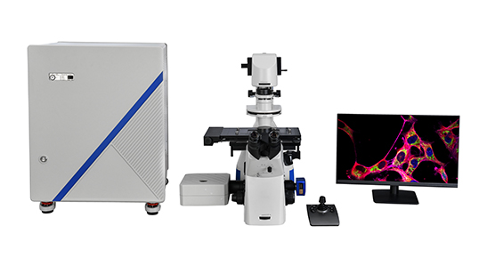
In 1957, Marvin Minsky proposed some basic principles of confocal microscope technology and developed the earliest prototype of confocal microscopy, which was awarded a patent in the United States.
However, at that time, there was a lack of applicable super-powerful light sources, and the data processing capability was not up to the required standard. In fact, this concept only stayed at the theoretical stage.
Until 1987, White and Amos published the article ” Confocal microscopy comes of age ” in the British “Nature”, marking that LSCM has become an important tool for scientific research.
Parts of the Confocal Microscopy: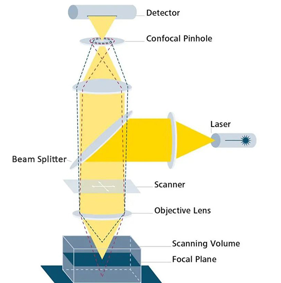
A: The laser
It can be chosen via a selection device and is matched with the fluorophores used in your experiment.
B: Beam splitter
It separates the excitation from the emitted light in the fluorescence beam path of the microscope.
C: Scanner
It guides them focused laser beam across the specimen, pixel-by-pixel, and line-by-line.
D: Objective lens
It determines the optical image formation and the resolution of the system.
E: Focal plane
This allows the user to focus the laser beam on any focal plane within the specimen.
The motorized Z-stepper allows us to move around the axial direction in small step sizes (approximately >10 nm) with high precision.
F: Confocal pinhole
Pinhole is a type of adjustable iris.
Pinhole allows the exclusion of most of the out-of-focus light from the acquired image and thus provides optical sectioning capacity.
The size of the pinhole can be set by using the software on the user’s computer.
G:Detector
It converts the photons into an electrical signal which is then used up by the computer to create an image of the specimen.
Principle of the Confocal Microscopy:
A confocal microscopy uses a laser light source, pinhole spatial filters, multiple objective lenses, and fluorescent dyes to generate a microscopic image. The laser provides excitation light, which reflects off of a mirror. The laser light then hits two mirrors that are mounted. These mirrors redirect the laser light, which effectively scans the sample and emits fluorescent light. The dye and the emitted light are then descanned by the same mirrors that originally scanned the excitation light.
From there, the emitted light passes through what’s known as a dichroic, a type of color filter used to selectively pass light of a small range of colors. This filtered light is directed toward the pinhole. The light passing through the pinhole is measured by a photomultiplier tube (PMT) or other types of photosensitive detectors, including photodiodes, and solid-state charge-coupled devices (CCDs). The pinhole filter eliminates anything that’s out of focus, producing a higher quality image than standard widefield microscopes.
The detector is attached to a computer that builds the image, one pixel at a time, as there is never a complete image of the sample. At any moment, only a single point of the sample is observed. The limitation of the confocal microscope is in the scanning mirrors.
The laser light intensity can be adjusted by neutral density filters. The scanning mirrors move quickly. One mirror tilts the laser beam in the X-direction, while the other tilts the laser beam in the Y direction. This is different from conventional microscopy in the sense that with a conventional microscope, light only travels as far as it can penetrate into the specimen. With confocal, it travels at a narrow depth, one level at a time, to achieve a limited, yet controlled depth of focus.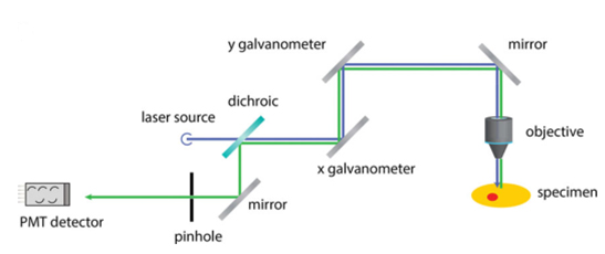
With the majority of software options, optical sections can be collected at the perpendicular lateral (x and y-axis) as well as transverse plans (x and z-axis) and the y and z-axis.)
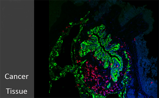
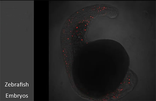
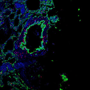
Applications of Confocal Microscopy:
In recent years confocal microscopy has been adapted into numerous fields – such as cell biology, life sciences, semiconductor inspection, materials science, neuroanatomy, and neurophysiology.
Key applications areas include:
- Stem cell research
- Photobleaching studies
- Fluorescence resonance energy transfer (FRET)
- Fluorescence recovery after photo-bleaching
- Fluorescence in-situ hybridization
- Lifetime imaging
- Multiphoton microscopy
- Total internal reflection fluorescence microscopy (TIRFM)
- DNA hybridization
- Membrane and ion probes
- Bioluminescent proteins
- Epitope tagging
![]()
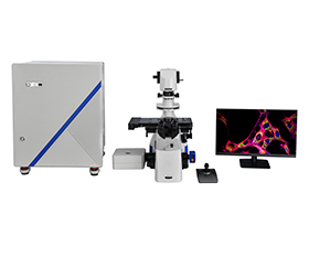
BestScope-BCF295 Laser Scanning Confocal Microscope
This confocal microscope can make a three-dimensional image of a translucent object through the moving lens system, and can accurately test the subcellular structure and dynamic process.
Feature:
- Infinite optical system
- Laser 405nm, 488nm, 561nm, 640nm
- Detector: 3 PMT. Wavelength: 400-750nm
- Confocal field of view: φ20mm inscribed square
- Transmitted Kohler illumination
The resources are collected and organized on the Internet, and are only used for learning and communication. If there is any infringement, please contact us to delete.