How to Choose a Camera for Fluorescence Microscope?
The camera is an essential component of any fluorescence microscope because it captures images of the specimen for observation. The fluorescence microscope uses fluorescent molecules to make objects visible. Fluorescent molecules are excited by light and emit light at a different wavelength. The emitted light is usually invisible to the human eye, but it can be detected by a camera and used to produce an image.
There are many factors that need to be taken into consideration when choosing a camera for fluorescence microscopes. These include:
Sensor Type: CMOS or CCD
All camera sensors are made up of an array of pixels which collect photons of light. Each pixel is composed of a photo-sensitive photodiode with silicon coupled to an electron storage well. When the photons hit the silicon substrate, photoelectrons are generated.
The electrons are stored in electron storage wells and then transferred (readout) to an amplifier. The amplifier reads the electrons and converts them to a voltage. Adjacent to the amplifier is an analog-digital (AD) converter which measures the voltage created by the electrons and translates this into a digital signal.
The more photons that hit the camera sensor, the more charge, and therefore signal, generated. The number of photons reaching the camera sensor is determined by the length of time exposed to light, the wavelength detected and the intensity of light.
The CMOS (complementary metal-oxide semiconductor) camera is one of the most popular types of sensors. They have a large field of view and high resolution. They are used for fluorescence microscopes due to fast frame rates, high dynamic range and low noise. In low-light environments, they can be used in conjunction with pixel binning to improve their photon collection.
In CMOS cameras, each pixel has its own amplifier, and one column of pixel connects to one AD converter. This results in CMOS cameras having a faster frame rate compared to CCD cameras. They also have a lower read noise, as each AD converter has more time to read the electrons, so can read them more slowly than the one AD converter in a CCD camera.
The CCD (charge coupled device) camera is another popular option, but they are more expensive than CMOS cameras and do not provide as high quality images as CMOS cameras do.
In CCD cameras, electrons in each pixel are passed along into the serial register. The serial registers pass electrons one at a time to the amplifier and AD converter, which translates the voltage into a signal.
As every pixel is read in the same way, by the same AD converter, the AD converter has a lot of pixels to read quickly. The faster the frame rate of the camera, the greater the uncertainty at the AD converter, therefore the greater the read noise of the camera.
Sensor Size:
Sensor size is also important as it effects the field of view. Most available microscope cameras use either 1/3, 1/2 or 2/3 inch sensors, with a few more expensive models having 1 inch. The larger sensor generally offers a wider field of view, and it also can capture more light and consequently better images it produces.
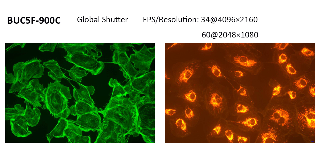
Global and Rolling Shutter:
CCD sensors have only one shutter type (global) while CMOS sensors are available in two types: rolling and global. Choosing the right sensor has a significant impact on image quality, especially when target objects are moving. In rolling shutter sensors, the pixels are exposed line after line. As a result, an object that has changed its position between signal capturing of two lines produces deviating image situations, generating space distortion in the image. A technical advantage of rolling shutter sensors is that they have fewer electronic parts in the pixel, which can result in less noise during readout. Meanwhile, global shutter sensors expose all pixels of the sensor at the same time. In this case, there is no time shift between the exposures of the different pixel lines, thereby generating no space distortions when objects are moving.
Quantum Efficiency (QE):
Quantum efficiency (QR) expresses the conversion ratio between photons of a given wavelength and electrons being freed on the image sensor to produce a signal. The higher the QE, the less light is required to fall on each pixel to obtain a usable signal. Peak QE denotes the wavelength at which the camera is best suited to convert photons into electrons. The camera’s overall QE graph indicates the camera’s QE in the range of each dye’s emission wavelength showing whether adequate conversion efficiency exists.
Frame Rate:
Frame rate is the number of frames per second that a camera can take. A higher frame rate means that the camera will be able to capture more detail in an image and reduce motion blur. For fluorescence microscopes, a frame rate of at least 60 frames per second is recommended.
CMOS cameras usually have a higher frame rate than CCD cameras. How quickly the camera signal is converted from an analog to digital signal also affects the frame rate, for example, a 16 bit sensor can transmit more data faster than an 8bit sensor.
Sensitivity and Signal-to-noise Ratio:
Sensitivity is the measure of how clear the camera can detect a fluorescent signal. It takes into account how much light the camera receives and how much light it generates. The higher the sensitivity, the more sensitive it is to fluorescent signals. A signal-to-noise ratio (SNR) is a measure of how strong the signal from fluorescent molecules are relative to background noise in an image. The higher SNR, the more sensitive it is to fluorescent signals and vice versa.
Binning:
Binning is used to increase the frame rate of a camera sensor. Electrons from adjacent pixels are combined in the serial register and readout together. E.g. in 2×2 pixel binning, information from 4 adjacent pixels are combined and readout as one ‘super pixel’. Binning increases the frame rate and signal-to-noise ratio but reduces spatial resolution.
Cooling System:
The temperature of the sensor has a central influence on the dark current, which worsens the SNR and image quality – especially when the light signals are weak and longer exposure times are needed. This means that cooling the cameras can be important, but it is not absolutely necessary in fluorescence imaging. Since cooling measures significantly impact the system costs, the majority of cameras aren’t actively cooled, which is already sufficient for applications with good fluorescence signals. But even in these cameras, the design influences the sensor temperature. Heat generation should be avoided by operating the camera with low power consumption.
Software:
The camera software usually covers most users’ needs including image saving, performing measurements, annotation and more. For fluorescence microscopes, the monochrome camera is a good choice because it can capture images in low light conditions and higher quantum efficiency. The black and white images can be colorized with software to reduces noise and eliminates artifacts while maintaining a fast frame rate.
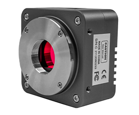
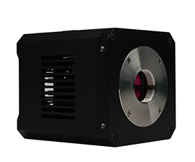
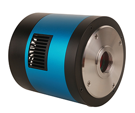
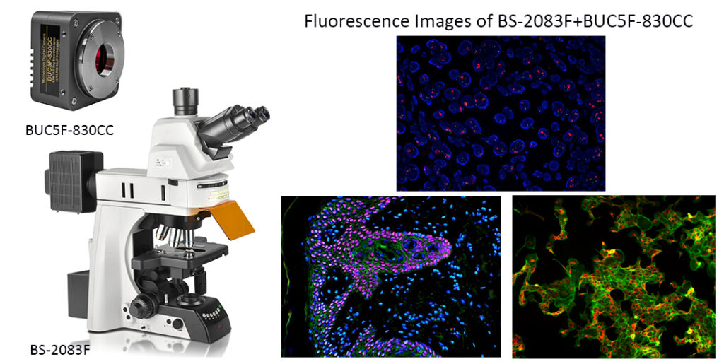
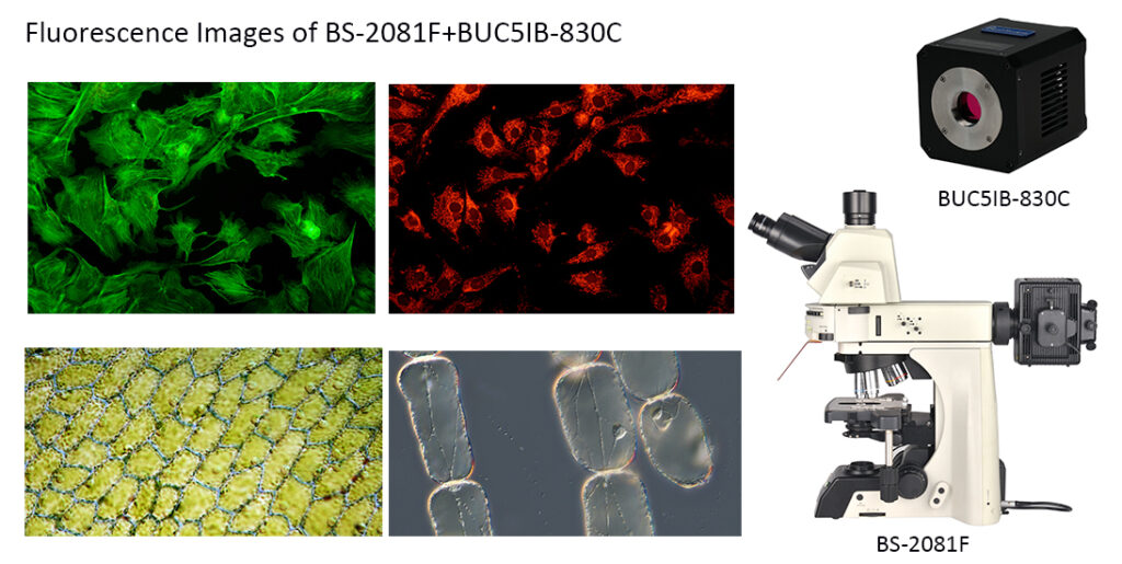
What camera does BestScope provide for fluorescence microscope?
|
Model |
Sensor & Size(mm) |
Pixel Size(μm) |
Frame Rate: |
Binning: |
Application |
|
8.3M/IMX485(Color) 1/1.2”(11.14×6.26) Rolling Shutter |
2.9×2.9 |
45@3840×2160 70@1920×1080 |
1×1 2×2 |
Fluorescent Light Environment |
|
|
8.3M/IMX485(Color) 1/1.2”(11.14×6.26) Rolling Shutter |
2.9×2.9 |
43@3840×2160 66@1920×1080 |
1×1 2×2 |
Low light Microscope Fluorescence Image |
|
|
6.0M/ICX694AQG(C) Rolling Shutter |
4.54×4.54 |
7.5@2748×2200 |
1×1 |
Low light Microscope Fluorescence Image |
The resources are collected and organized on the Internet, and are only used for learning and communication. If there is any infringement, please contact us to delete.