What are the Applications of Microscopes in Biology?
 Microscopes have been a crucial tool in the field of biology for centuries. With the advancement of technology, microscopes have become even more useful in various branches of biology. In this article, we will discuss the different applications of microscopes in botany, human biology, zoology, and microbiology.
The Application of Microscopes in Botany:
One of the application of microscopes in botany is the study of plant cells. Microscopes allow scientists to observe the various structures of plant cells, such as the cell wall, nucleus, mitochondria, chloroplasts, and other organelles. By observing these structures, scientists can understand how they function and how they contribute to the overall health and growth of the plant.
Another application of microscopes in botany is the study of plant tissues. Microscopes allow scientists to observe the structure and arrangement of cells in different tissues, such as the epidermis, cortex, and vascular tissue. By studying the arrangement of cells in tissues, scientists can understand how plants transport water and nutrients, how they respond to environmental stresses, and how they grow and develop.
Microscopes also play a vital role in the study of plant pathogens. By observing the structure and behavior of plant pathogens, scientists can develop strategies to control and prevent plant diseases. Microscopes allow scientists to see the details of plant pathogens, such as bacteria, fungi, and viruses, that are invisible to the naked eye.
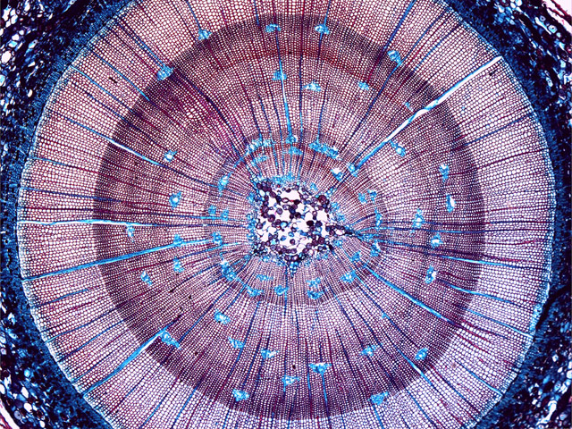
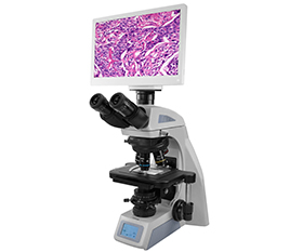
BS-2085 Biological Microscope Pine Sample

Leaves under Stereo Microscope
The Application of Microscopes in Zoology:
In zoology, microscopes are used to study a wide range of organisms, from bacteria to insects to larger animals. They are used to observe the structure and function of cells, tissues, and organs. Microscopes are also used to study the behavior of animals, such as the movement of cells during development or the interactions between different species.
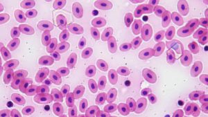
Frog Blood Smear (Taken By BS-2074T 40x Objecvtive+BHC4-1080P8MPB)
The Application of Microscopes in Human Biology:
In human biology, one of the most common uses is to examine blood cells. By placing a drop of blood on a slide and using a microscope, scientists can observe the different types of blood cells, such as red blood cells, white blood cells, and platelets. This helps diagnose various diseases and conditions, such as anemia, leukemia, and infections.
Microscopes are also used to examine tissue samples. Biopsies, which involve removing a small piece of tissue for examination, are commonly used to diagnose cancer and other diseases. By using a microscope, pathologists can examine the tissue sample and identify any abnormalities or cancerous cells.
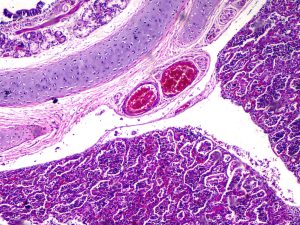
Interstitial Pneumonia (Taken By BUC2D-200C)
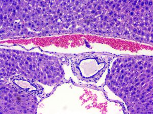
Liver (Taken By BUC2D-1200C)
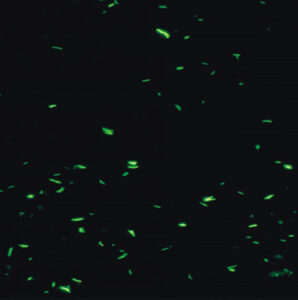
The Application of Microscopes in Microbiology:
Optical Microscopes can magnify objects up to 1000 times their actual size, making them ideal for studying bacteria, fungi, and other microorganisms.
The most common application of microscopy in microbiology is to observe the morphology or physical properties of microorganisms. Observing the shape, size, and arrangement of cells can provide valuable information for the identification and classification of microorganisms.
Microscopes are also used in microbiology to identify microbial pathogens. Pathogens are microorganisms that can cause disease in humans and animals. Identifying these microbes is critical for the diagnosis and treatment of infectious diseases.
BS-2044 Tuberculosis Sample
Electron microscopes, on the other hand, use a beam of electrons to illuminate the sample and magnify the image. They can magnify objects up to 100,000 times their actual size, allowing scientists to observe the structure of viruses and other small microorganisms in great detail. However, electron microscopes require expensive equipment, specialized training, and can only be used to observe dead, stained samples.
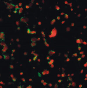
Confocal microscopes use lasers to illuminate the sample and create a 3D image. They are particularly useful for studying the behavior of microorganisms in real-time and can be used to observe live, unstained samples. Confocal microscopes are also used to study the internal structure of cells and tissues.
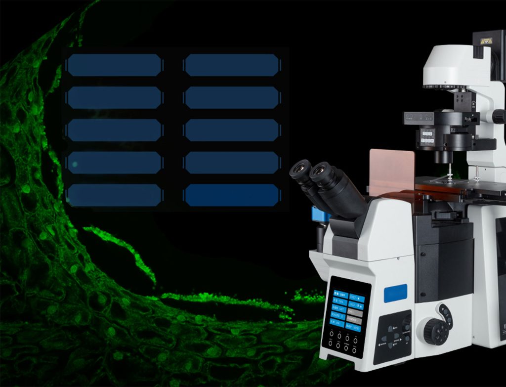
BCF297 Laser Scanning Confocal Microscopy
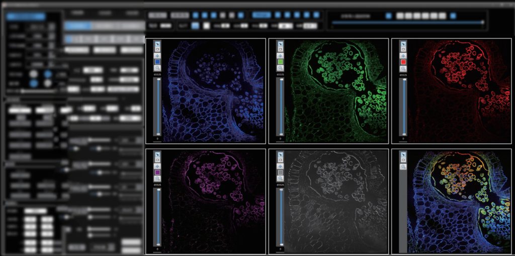
BCF297 Software
Overall, if you are looking for a microscope that offers optimal imaging capabilities, powerful magnification, and a range of useful applications for biological research, please feel free to contact us.
The resources are collected and organized on the Internet, and are only used for learning and communication. If there is any infringement, please contact us to delete.