


BS-2098 Research Inverted Microscope
Introduction
BS-2098 fully automatic research inverted microscope integrates multiple technologies such as electrical control, fluorescence, phase contrast, relief phase contrast, simple polarization, DIC, and objective lens with correction ring, achieving a three-level electric control of a single machine, TPC, and computer.
Details
Overview
Packaging & Delivery
Packaging Details:Strong Carton with Polyfoam Protection
Port:Beijing
Lead Time:Within 2-4 Weeks after Receiving Payment
Introduction
As a representative of Chinese optical, mechanical, electrical and computing integrated microscope, BS-2098 fully automatic research inverted microscope integrates multiple technologies such as electrical control, fluorescence, phase contrast, relief phase contrast, simple polarization, DIC, and objective lens with correction ring. A three-level electric control of single machine, TPC and computer is realized. BS-2098 has powerful expansion functions, providing a more complete solution for life science research.
With flexible structural design and excellent optical performance, BS-2098 can provide you with high-definition wide-field images, providing powerful technical support for cell observation, single-cell patch clamp, microinjection and other research and applications.
Features
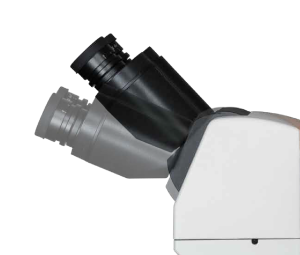
1.Elevation adjustable viewing head
BS-2098 is equipped with a 20-45° adjustable binocular viewing head, which can raise the eye point by 78mm (65mm interpupillary distance) as required. It can be observed easily and quickly even in a standing state, which is effective avoiding fatigue of eyes and limbs.
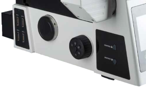
2.Integrated button
BS-2098 retains the traditional coarse and fine adjustment mode, cancels the gear mechanism, integrates electric control technology, and realizes manual and automatic integration. The brightness, objectives, attenuator turntable and fluorescent turntable can be quickly switched or rotated through the corresponding buttons on the button panels on both sides. AF one-key auto focus can quickly adjust the Z-axis height according to the real-time image, and eliminates the need for fine-adjusting steps and improves work efficiency.
3.Low-position platform handwheel
The low-position platform handwheel can be rotated 360°, which can effectively reduce the hand fatigue caused by long-term operation and is ergonomic and improves the convenience of operation.
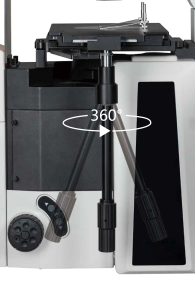
4.Tilting transmission lighting structure
The tilting structure of the transmission bracket ensures a large working space for users and facilitates the replacement of specimens.
![]()
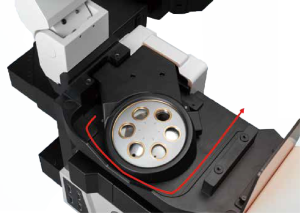
5.Waste liquid drainage structure
The drainage groove structure is designed under the objective nosepiece, which can prevent the optical components and modules from accidentally being wetted by the cell culture medium or soaking liquid, contaminating and damaging the microscope, simplifying maintenance.
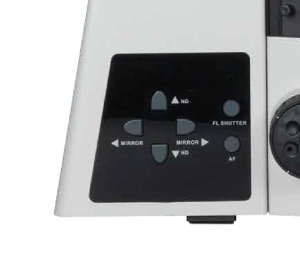
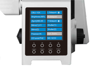
6.Integrated panel control
BS-2098 front digital display screen can display the status of multiple components such as microscope objective lens magnification, brightness, and fluorescence band in real time, and can complete the setting of all observation methods, the selection of optical port status, and the switch of fluorescence shutter. Greatly enhance the customer experience and make research work more convenient.
7.High scalability
BS-2098 has a very large rack that provides redundant space for third-party configurations. You can choose single-layer or double-layer optical path according to your needs, up to 16 color filters can be replaced. Multi-light port design can be equipped with confocal module, wide-field fluorescent camera, infrared camera, etc., providing maximum scalability for the research work.
8.Software
With 50 different image adjustment and processing algorithms, the software is able for photography, image stitching, auto focusing, DOF extension, multichannel acquisition, image measurement. One-click to generate a report, reliable and effective.
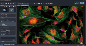
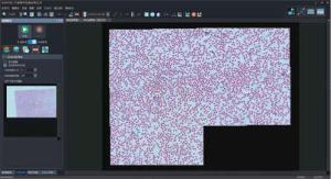
Image stitching
It is able to stitch the local image, with 8*8 field of view.
Cell measurement
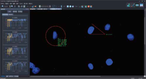
Image software with measuring function, is available to analyze cell size, intercellular space, synapse length and so on, including distance, angle, rectangle and round measurement.
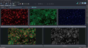
Fluorescent synthesis
The image from different fluorescent channels, are able to be synthesized automatically and kept separately. Each image is available to be adjusted and the final composited image would be auto updated.
Report generation
Built-in module to fast generate a report, supports to output WORD document.
Specification
|
Item |
Specification |
BS-2098 |
|
|
Optical System |
Infinity Color Correction Optical System |
● |
|
|
Viewing Head |
20-45 degree tilting binocular tube, interpupillary distance: 50-76mm |
● |
|
|
Eyepiece |
High eye point wide field plan eyepiece PL10X/22mm, with adjustable diopter |
● |
|
|
Objectives |
Long Working Distance Plan Semi- Apochromatic Objectives |
4X/NA=0.13, WD=17mm |
○ |
|
10X/NA=0.3, WD=8.8mm |
○ |
||
|
20X/NA=0.45, WD=6.5-7.6mm, coverslip thickness: 0-2mm |
○ |
||
|
40X/NA=0.6, WD=2.85-4.05mm, coverslip thickness: 0-2mm |
○ |
||
|
60X/NA=0.7, WD=1.42-2.1mm, coverslip thickness: 0-1.3mm |
○ |
||
|
Long Working Distance Plan Semi- Apochromatic Positive Phase Contrast Objectives |
4X/NA=0.13, WD=17mm |
● |
|
|
10X/NA=0.3, WD=8.8mm |
● |
||
|
20X/NA=0.45, WD=6.5-7.6mm, coverslip thickness: 0-2mm |
● |
||
|
40X/NA=0.6, WD=2.85-4.05mm, coverslip thickness: 0-2mm |
● |
||
|
60X/NA=0.7, WD=1.42-2.1mm, coverslip thickness: 0-1.3mm |
● |
||
|
Infinity Plan Apochromatic Objectives |
4X/NA=0.16, WD=12.8mm |
○ |
|
|
10X/NA=0.4, WD=3.2mm |
○ |
||
|
20X/NA=0.75, WD=0.6mm |
○ |
||
|
40X/NA=0.95, WD=0.15mm |
○ |
||
|
60X/NA=0.9, WD=0.26mm |
○ |
||
|
(Oil) 60X/NA=1.25, WD=0.14mm |
○ |
||
|
(Oil) 100X/NA=1.35, WD=0.13mm |
○ |
||
|
Infinity Plan Super Apochromatic Objectives |
10X/NA=0.4, WD=3.1mm |
○ |
|
|
20X/NA=0.8, WD=0.6mm |
○ |
||
|
40X/NA=0.95, WD=0.18mm |
○ |
||
|
(Oil) 60X/NA=1.42, WD=0.17mm |
○ |
||
|
(Oil) 100X/NA=1.45, WD=0.13mm |
○ |
||
|
Long Working Distance Plan Semi-Apochromatic Relief Phase Contrast Objectives |
10X/NA=0.3, WD=8.8mm |
○ |
|
|
20X/NA=0.45, WD=6.5-7.6mm, coverslip thickness: 0-2mm |
○ |
||
|
40X/NA=0.6, WD=2.85-4.05mm, coverslip thickness: 0-2mm |
○ |
||
|
Microscope Body & Nosepiece |
Low position coarse and fine coaxial electric focusing mechanism, range: 10.5mm, precision: 1μm. Built-in electric upper camera port, splitting ratio: 100:0 / 0:100. Built-in electric left camera port, splitting ratio: 0:100 / 50:50 / 100:0, dual optical path, with fluorescent light barrier. Electric bright field sextuple nosepiece with DIC slot and upper optical port CTV adapter. |
● |
|
|
Base mounting bracket |
○ |
||
|
Right camera port, splitting ratio: 100:0 / 0:100, field of view: 16mm. Built-in 1X CTV, C-mount adapter |
○ |
||
|
Illumination |
Pillar tilt mechanism, Koehler transmission illuminator, adjustable condenser holder with 65mm stroke. 4 filters holders with LBD, Green filter, Neutral filter for halogen models or Neutral filter for LED models. |
● |
|
|
12V/100W halogen illumination, filament center preset |
● |
||
|
12V/100W halogen lamp |
● |
||
|
10W cool color LED light illumination, color temperature 5000K |
○ |
||
|
Condenser & Iris |
Electric septuple condenser, NA 0.55, WD=27mm. 3 holds for Φ30mm (phase contrast), 4 holds for Φ38mm (DIC), support for bright field/phase contrast/DIC (with polarizing kit) (BS20980036) |
● |
|
|
Cable 100cm |
● |
||
|
4X Phase contrast annular iris (for BS20980036) |
● |
||
|
10X Phase contrast annular iris (for BS20980036) |
● |
||
|
20X/40X/60X Phase contrast annular iris (for BS20980036) |
● |
||
|
Super long working distance manual condenser with 5 holes, NA 0.3, WD=73mm, support for 4X-60X phase contrast, simple polarizing observation and 10X-40X relief phase contrast observation. (BS20980041) |
○ |
||
|
4X Phase contrast annular iris (for BS20980041) |
○ |
||
|
10X Phase contrast annular iris (for BS20980041) |
○ |
||
|
20X/40X/60X Phase contrast annular iris (for BS20980041) |
○ |
||
|
10X Oblique iris (for BS20980041) for relief phase contrast observation |
○ |
||
|
20X Oblique iris (for BS20980041) for relief phase contrast observation |
○ |
||
|
40X Oblique iris (for BS20980041) for relief phase contrast observation |
○ |
||
|
360 degree rotatable polarizer (for BS20980041), mandatory for simple polarizing and relief phase contrast observation |
○ |
||
|
DIC |
Transmitted DIC kit |
○ |
|
|
10X transmitted DIC ring (for BS20980036) |
○ |
||
|
20X transmitted DIC ring (for BS20980036) |
○ |
||
|
40X/60X transmitted DIC ring (for BS20980036) |
○ |
||
|
Analyzer kit (for BS20980036) |
○ |
||
|
Fluorescent Module |
Fluorescence attachment with 8 holes, with electric shutter |
● |
|
|
Cable 20cm |
● |
||
|
Dust cap |
● |
||
|
B fluorescence filter cube |
● |
||
|
G fluorescence filter cube |
● |
||
|
UV fluorescence filter cube |
● |
||
|
L type fluorescence attachment, with filter plugboard, ND25 attenuation piece, ND50 attenuation piece |
● |
||
|
100W mercury lamp house |
● |
||
|
Fluorescence power supply |
● |
||
|
100W DC mercury lamp |
● |
||
|
Electric attenuation holder with 5 holes, with bright field/light barrier/ ND6/ND25/ND50 (no need to use extension accessory) (BS20980064) |
○ |
||
|
Cable 100cm (mandatory for BS20980064) |
○ |
||
|
Extension accessory (must be chosen when BS20980064 is not selected) |
● |
||
|
Stage |
Manual mechanical stage, size: 300mm(X)*240mm(Y), moving range: 135mm(X)*85mm(Y), stage thickness: 30mm. Right universal handle, X/Y axis limitable and lockable, moving range 50mm * 50mm after locked; with pressure clap for holding slices and culture flasks, with Φ110mm replaceable disc (inner Φ30), with metal stage plate with waist shaped hole. |
● |
|
|
Marzhauser electric stage, moving range: 120mm(X)*80mm(Y), resolution: 0.02μm, repeatability: 1μm, with control box and operating handle, with slide holder. |
○ |
||
|
Φ36 petri dish holder (for Marzhauser electric stage) |
○ |
||
|
96 holes petri dish holder (for Marzhauser electric stage) |
○ |
||
|
Terasaki holder (for Marzhauser electric stage) |
○ |
||
|
PRIOR electric stage, moving stage:114mm(X)*75mm(Y), resolution: 0.01um, two-way, repeatability: 0.2um, with controller, electric control box and cable, with slide holder |
○ |
||
|
Φ36 petri dish holder, with adjustable petri dish sample holder (for PRIOR electric stage) |
○ |
||
|
96 holes petri dish holder, recessed porous plate sample holder, suitable for 85*128mm orifice plate |
○ |
||
|
Terasaki holder, concave cells with bottle sample holder, suitable for 85*128mm culture flask |
○ |
||
|
Electric Control Box & PC |
Electric control box, input voltage 90-265VAC wide voltage, output 12V100W or 12V10W.Digitally adjustable output voltage through CAN, in addition to three outputs of 24V5A/15V5A/5V5A, equipped with forced air cooling, including one 3C power cord. |
● |
|
|
DB26 Cable 200cm, connecting the electric control box to the frame |
● |
||
|
7-inch LCD display controller, capable of operating the electronic control components of the body, with a 12V 5A medical adapter, including DB9 external cable. |
○ |
||
|
DELL PRECISIOM T3650 (I7 11700, 16GB/256GB+1TB/P620) +P2722H |
○ |
||
|
DB9 Cable 200cm, connecting the PC to the electric control box |
● |
||
|
USB-CAN card. When the customer purchases computer by themselves and requires computer to achieve microscope electric control, it must be paired. |
● |
||
|
PCIE-CAN card. When the customer purchases computer by themselves and requires computer to achieve microscope electric control, it must be paired. |
○ |
||
|
Telescope |
Telescope (Φ30) |
● |
|
|
C-mount Adapter |
0.5X C-mount adapter, adjustable focus |
○ |
|
|
0.65X C-mount adapter, adjustable focus |
● |
||
|
1X C-mount adapter, adjustable focus |
○ |
||
|
Other Accessories |
Fluorescent free oil 30ml |
○ |
|
|
Internal hexagonal Spanner M3 for phase contrast adjusting screw |
● |
||
|
Internal hexagonal Spanner M4 |
● |
||
|
Internal hexagonal Spanner M5 |
● |
||
Note: ● Standard Outfit, ○ Optional
Application

Cell observation
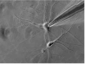
Single-cell patch clamp
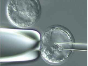
Microinjection and other research and applications
Accessories
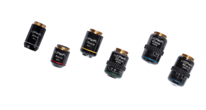
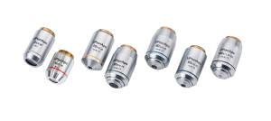
1.Long Working Distance Semi-Apochromat Objectives
A complete set of long working distance semi-apochromatic objective lenses has been developed for cell observation. The objective lens with correction rings have a strong advantage in observing glass substrates and petri dished of different thicknesses, achieving precise focusing by correcting coverage differences.
Long Working Distance Plan Semi-Apochromatic Objectives
Long Working Distance Plan Semi-Apochromatic Positive Phase Contrast Objectives
Long Working Distance Plan Semi-Apochromatic Relief Phase Contrast Objectives
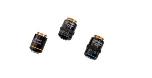
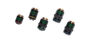
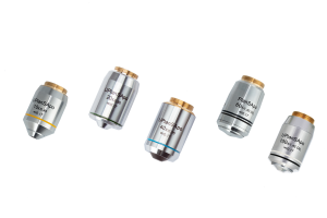
2.Infinite Plan Apochromatic Objectives
3.Infinite Plan Super Apochromatic Objectives
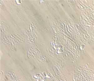
Sample Image
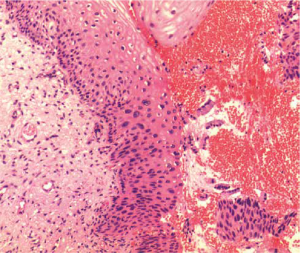
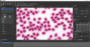
Skin papilloma (Bright field / 40X)
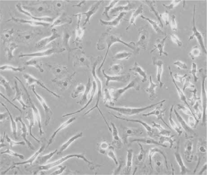
Normal rat kidney cells (Phase contrast /10X)
Oral epithelial cells (Relief phase contrast / 10X)
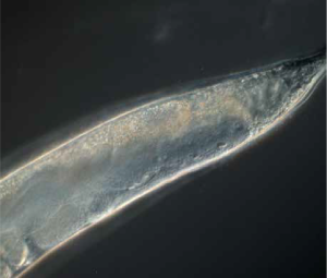
Nematode (DIC / 40X)
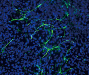
Human smooth muscle cells (Fluorescence / 20X)


")
")


