Karyotype Analysis Microscopy Instrument Solution
 What’s Karyotype Analysis
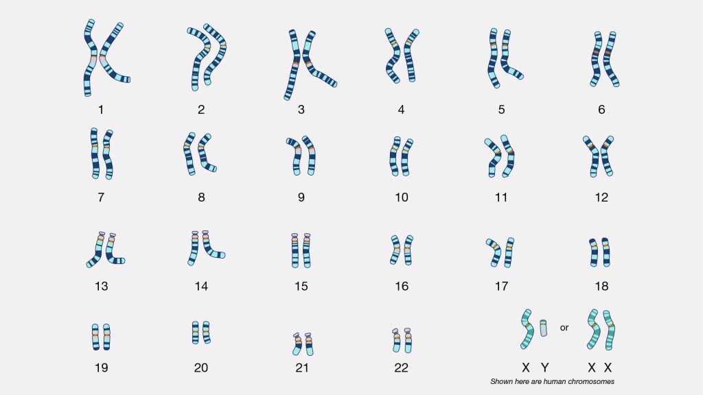
Karyotype refers to the phenotype of chromosomes in the metaphase of mitosis, which is a summary of the number, size and morphological characteristics of chromosomes. Chromosomes of different species all have their own specific structural and morphological characteristics, including the number of chromosomes, length, centromere, arm ratio, satellite size and so on. These morphological features are relatively stable. Abnormal karyotypes of chromosome can serve as key indicators for diagnosing various genetic disorders.
Karyotype analysis is an examination of the characteristics of chromosomes in somatic cells, such as length, centromeric position, arm ratio and satellite size. The research object is the chromosomes in the metaphase of somatic cell division. Karyotype analysis has great significance for exploring the mechanisms of genetic diseases in humans and other animals, species kinship and the identification of distant hybrids.
The Application of Karyotype Analysis
Some diseases are associated with abnormal chromosomal structure or number. The following are some examples of the relationship between common chromosomal abnormality genetic diseases in humans and karyotypes:
Down syndrome, mainly presents with low intelligence, distinctive facial features, etc., and the karyotypes of it is an extra chromosome 21.
Cri du chat syndrome is clinically characterized by crying like a cat, low intelligence, etc., and karyotype shows partial missing of the short arm of chromosome 5.
When the two ends of a chromosome break and connect to form a ring, it may lead to developmental delay or deformity.
One parent carrying a balanced translocation or other structurally abnormal chromosome may lead to repeated miscarriages or fetal arrest
Other diseases like hematological diseases may also be related to chromosomal abnormalities. For instance, 95% of patients with chronic myeloid leukemia (CML) have a translocation of the Philadelphia chromosome, which is the translocation of chromosomes 9 and 22, forming the BCR-ABL fusion gene.
Therefore, karyotype analysis is an important diagnostic tool for genetic diseases, reproductive abnormalities and certain hematological disorders.
The methods of Karyotype Analysis
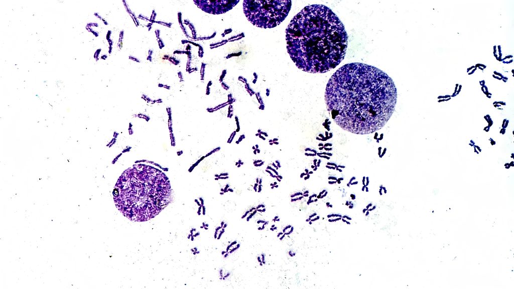
The sample for karyotype analysis include peripheral blood, bone marrow cells, skin and amniotic fluid, etc. Traditional karyotype analysis of chromosomes takes metaphase chromosomes as the research object. Colchicine is needed to make the sample cells maintain in the metaphase to obtain a sufficient amount of metaphase cells. After a series of options such as low permeability, fixation and slide making, stain the sample with the dyes such as acetic acid magenta or gentian violet. Then observe the samples under an optical microscope to determine whether there are any abnormalities in the number and structure of chromosomes to assess the risk of a certain disease. This is a non-banding karyotype analysis.
On the other side, chromosome banding karyotype analysis, processing chromosome samples with banding techniques to make the chromosomes display a certain number of bands of varying degrees of coloration and different widths along the longitudinal axis, with alternating light and dark patterns. Each chromosome has a unique and fixed band pattern, mainly depending on the interaction among DNA, proteins and dyes. Different banding techniques result in different band patterns displayed on chromosomes.
Common chromosome banding techniques include G-banding, Q-banding, R-banding and C-banding, etc. Q-banding technique, uses mustard quinacrine or quinacrine dihydrochloride to show fluorescent band patterns. G-banding technique, uses trypsin digest the chromosome sample and then stained with Giemsa to form bands. The G-banding looks similar with Q-banding, but G-banding staining is not limited by the short fluorescence time like Q-banding staining and G-banding can be observed under an ordinary biological microscope not a fluorescence microscope. The specimens also can be preserved for a long time, so it is widely used. In addition, there are R-banding that can produce the opposite pattern of G-banding, and banding techniques show special chromosomal structures, such as C-banding showing structural heterochromatin, T-banding showing telomeres, and NORs (nucleolar organizing regions).
Karyotype Analysis Microscopy Instrument Solution
Chromosome karyotype analysis requires the use of a biological microscope for observation, mainly to identify whether there are any abnormalities in the number or structure of chromosomes. Generally, the diameter of human chromosomes is 0.2-2μm, and the length variation range is 0.2-50μm micrometers. In karyotype analysis, the chromosomes to be observed should be in the metaphase of mitosis. In the metaphase of mitosis, a pair of chromosomes are connected by centromeres, and the distance between the two chromosomes is relatively close. Therefore, high-resolution high magnification microscope objectives are needed. Low-resolution image may affect the judgment of chromosome morphology. High numerical aperture generally represents higher resolution. With a 40x objective and a 100x oil immersion objective with high N.A., observers can easily distinguish the number and structure of chromosomes.
Due to the limitations of sample preparation technology, chromosomes may be located at different focal planes. So, a fine focusing system with high precision is needed. High-resolution focusing system can get clear images of chromosomes more conveniently and quickly. Also, the wide-field eyepieces, the white LED light source with adjustable brightness, the mechanical stage with low hand position adjustment, the coaxial coarse and fine focusing system with low hand position adjustment and the ergonomically designed microscope body can make the process of chromosome karyotype analysis for users much easier.
If karyotype analysis such as Q-banding that requires fluorescence coloration is to be conducted, a fluorescence microscope should be used. To improve efficiency, a motorized biological microscope can be used. Especially for motorized biological microscopes with digital section scanning systems, they not only increase efficiency but also prevent duplicate counting or omission caused by human factors.
In addition to manual analysis, specialized karyotype analysis software can be used to automatically and intelligently count and calculate the lengths of long and short arms, etc., to screen for abnormal chromosomes. However, due to the different banding characteristics of chromosomes, even with the use of specialized software, researchers still need to verify them manually and draw conclusions after analyzing and comparing multiple groups.
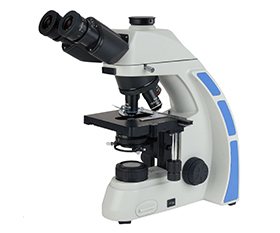
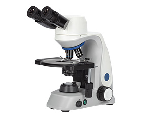
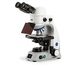
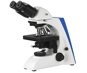
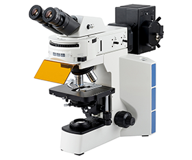
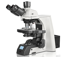
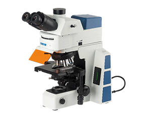
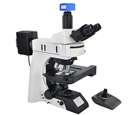
Recommended Biological Microscope
BS-2044 Series Biological Microscope
BS-2047 Series Biological Microscope
BS-2048 Series Biological Microscope
BS-2063 Series Biological Microscope
BS-2064 Series Biological Microscope
BS-2081 Series Research Biological Microscope
BS-2082 Series Research Biological Microscope
BS-2084 Series Biological Microscope
BS-2085 Series Motorized Automatic Biological Microscope
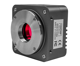
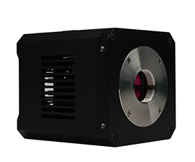
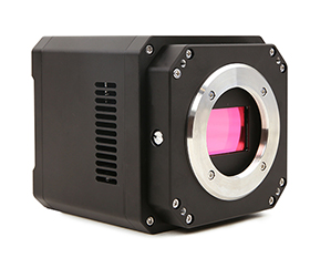
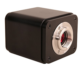
Recommended Microscope Digital Camera
BUC5F Series C-mount USB3.0 CMOS Camera (HISPVP)
BUC5IB Series Cooled C-mount USB3.0 CMOS Camera
BUC5IC Series TE-Cooling M52/C-mount USB3.0 CMOS Camera
BWHC4-4K Series HDMI/NETWORK/USB Multi-outputs C-mount CMOS Camera