Rabies Microscopy Instrument Solution
 What’s Rabies
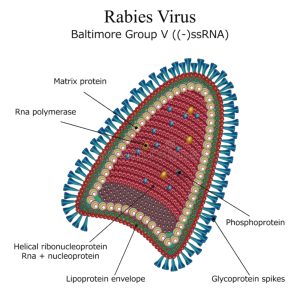
Rabies is an acute zoonotic infectious disease mainly affecting the central nervous system, caused by the rabies virus. The clinical manifestations include specific hydrophobia, fear of wind, restlessness, pharyngeal muscle spasm, progressive paralysis, etc. Rabies virus is usually transmitted from sick animals to humans through saliva from bites. The rabies virus is mainly transmitted among animals, such as dogs, cats, foxes and bats, etc. which may all fall ill and infect others. In modern life, stray dogs are the main source of animal infection.
If people are bitten by a sick dog and left untreated, about 30% to 70% of them will be infected with rabies and develop the disease after the incubation period. The incubation period of human rabies ranges from 10 days to over one year, usually 30 to 90 days. Those who have been bitten on the head, neck or hands have a shorter incubation period. During the incubation period, the infected person may show no symptoms at all. Rabies patients usually die within 3 to 10 days after the onset of the disease. The mortality rate after the onset of the disease was nearly 100%, with only one case of recovery in 1971.
If appropriate treatment is given immediately after being bitten by a rabies animal, such as debridement and vaccination, it is rare to develop the rabies. Before and after exposure or infection, vaccination is the only effective way to fight the disease, because once symptoms appear, no treatment can work.
Diagnosis of Rabies
If there are typical symptoms such as fear of water or pharyngeal spasm, it is not difficult to diagnose. In the incubation period, with no symptom or mild symptom, it is generally not easy to diagnose. Asking if there is a history of being bitten or if there are any abnormal sensations at the wound can help with diagnosis.
Clinical diagnosis
Similar to common viral encephalitis, the most specific and diagnostically significant feature is the presence of Negri bodies, eosinophilic inclusions that exist in the cytoplasm of nerve cells in 80% of rabies patients. The Negri bodies are round or oval in shape, about 3-10μm in size, with neat edges and 1-2 small dots resembling cell nuclei inside. Negri bodies are the colonies of the virus. Under and electron microscope, rod-shaped virus can be seen in the Negri bodies. If Negri bodies are found in the human brain or animal brain cells, a definite diagnosis of Rabies can be made.
Laboratory diagnosis
There are serval methods in laboratory diagnosis, including Negri bodies examination in brain tissue, immunofluorescence detection of antibodies, nucleic acid testing, animal inoculation experiment with secretions, viral RNA examination with reverse transcription PCR, serological antibody test, etc.
Microscopy Instrument Solution of Rabies
Fluorescence microscope
Antibodies or antigens can be detected by immunofluorescence. For samples with positive antigen or nucleic acid test results, it is also possible to conduct immunofluorescence detection after virus isolation.
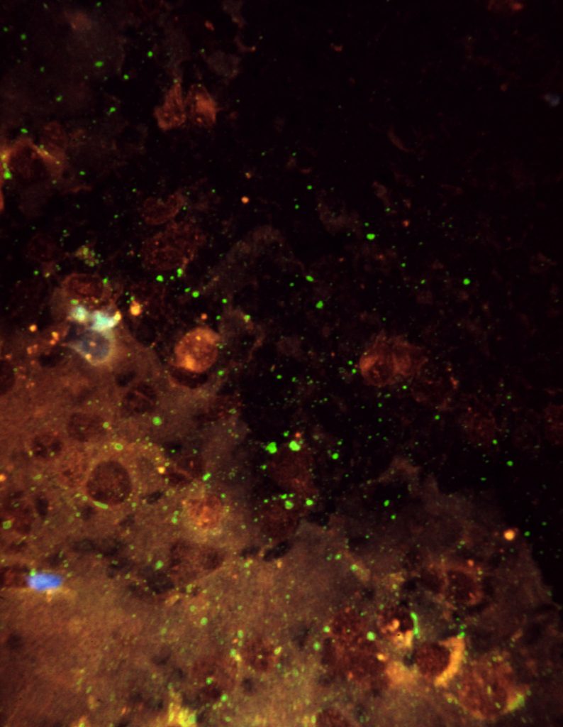
Micrograph from F. A. Murphy
Immunofluorescence detection is based on the principle of antigen-antibody reactions. First, a known antigen or antibody is labeled with a fluorescent group. And then this antibody or antigen with fluorescent is used as a probe to detect the corresponding antigen or antibody within cells or tissues. The fluorescence microscopes can be used to observe the cells or tissues where fluorescence is located, to determine the nature and location of antigens or antibodies.
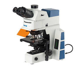
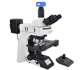
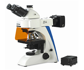
Recommended Fluorescence Microscope
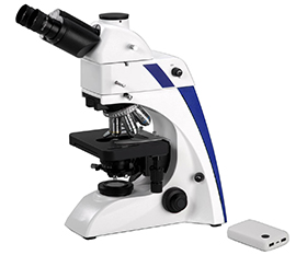
BS-2063F(LED) LED Fluorescence Microscope
BS-2063F2(LED) LED Fluorescence Microscope
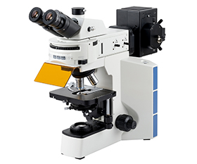
BS-2064F Fluorescent Biological Microscope
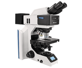
BS-2076F Research Fluorescent Biological Microscope
BS-2081F Research Fluorescent Biological Microscope
BS-2082F Research Fluorescent Biological Microscope
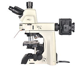
BS-2085F Motorized Fluorescent Biological Microscope
BS-2091F Inverted Fluorescent Biological Microscope
BS-2097 Research Inverted Microscope
BS-2098 Research Inverted Microscope
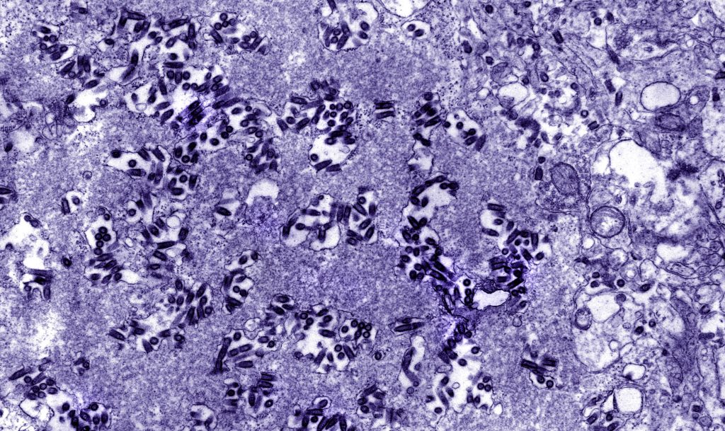
Electron microscope
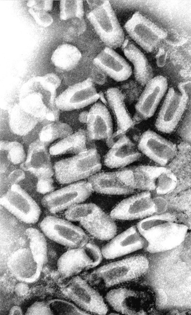
Micrograph from F. A. Murphy
Electron microscopy examination can provide a rapid and intuitive diagnosis for the morphological identification of rabies virus. Under a scanning electron microscope, it can be observed that the rabies virus can undergo agglutination reactions with goose red blood cells and adhere to the surface of goose red blood cells. Under a transmission electron microscope, rabies virus particles appear significantly bullet-shaped.
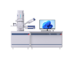
Recommended Electron Microscope
BSEM-691 Tungsten Filament Scanning Electron Microscope
BSEM-801 Field Emission Gun Scanning Electron Microscope
BSEM-810 Field Emission Gun Scanning Electron Microscope
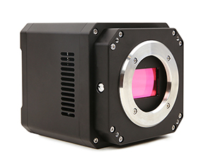
Recommended Microscope Digital Camera
BUC5F Series C-mount USB3.0 CMOS Camera (HISPVP)