
What is Dark Field Microscopy Used for?
Dark field is a lighting technique of oblique illumination used in microscope observation. With dark field condenser and special objectives, it can achieve a bright sample image with a black background. When the bright field illumination cannot provide sufficient contrast for observation, dark field illumination can be considered. Let’s have a detailed look at the principle, advantages and disadvantages, component of dark field and what is dark field microscopy used for.

Principle – How the Dark Field works
In the bright field microscopy, the condenser focuses the light directly through the sample and into the objective. The contrast of the image depends on the light absorbed by the sample. It works well for naturally colorful or artificially stained samples, but has poor imaging results for transparent or unstained low-contrast samples.
The working principle of dark field microscopy is to block the central light and allow oblique illumination to illuminate the sample. Only the scattered, diffracted and refracted light enters the objective, thus obtaining an image of a bright sample in a dark background.
The dark field attachment is an opaque shading plate that blocks the central light. The dark field condenser causes the light emitted from the surface to form an inverted hollow light cone, focusing on the sample plane. The oblique light that shines on the sample, such as the cell membrane or cell organelle, will undergo diffraction, reflection and refraction and then enter the objective, while the other empty areas appear dark in the images because the oblique light that shines on the empty areas will just intersect each other and miss the objective. Eventually, an image of a bright and vivid sample in a dark background is done.
To prevent the oblique light from directly entering the objective, the NA value, numerical aperture, of the objective needs to be lower than that of the condenser. Otherwise, the oblique light emitted by the dark field condenser within the receiving angle of the objective will directly enter the objective, and the strong light entering the eyepiece will prevent the sample from being observed.
Advantages & Disadvantages – Why Choose the Dark Field
Advantages – High contrast imaging
Dark field microscopy imaging has a high contrast, suitable for observing transparent samples or samples with low contrast that are difficult to observe in bright field microscopy.
Advantages – No need for sample staining
The samples can be easily observed without the need for complex staining procedures.
Advantages – Suitable for live samples
Since samples can be observed without staining, the live aquatic organisms and living cells can be directly observed in their nature state.
Disadvantages – Require high lighting
Since most of the light from the illuminator is blocked in dark field microscopy, a large amount of light is required. Otherwise, the field of view will be extremely dark. So, a high-intensity illuminator is needed for the dark field microscopy. For living specimens that are sensitive to light, long-term exposure to strong light may cause photodamage or photobleaching.
Disadvantages – Limited detail display
The display of internal details of the sample is limited in dark field microscopy. If the transparent sample has complex or dense internal structures, the dense structures will cause the scattered light to overlap, resulting in a blurry halo, which will affect the observation. At the same time, due to scattering, impurities on the slide, such as dust and debris, will be very obvious, reducing image quality.
Disadvantages – NA value limitation
Since the NA value of the objectives must be lower than that of the condenser, for oil-immersed objective with high NA value, oil-immersed condenser with high NA value must be used, which increases cost and operation complexity. At the same time, the condenser needs to be precisely calibrated before use to ensure uniform illumination within the field of view.
The comparison between bright field and dark field is as follows.
|
Bright Field |
Dark Field |
|
|
Image Background |
Bright |
Dark |
|
Whether the Sample Needs Staining |
The sample usually requires staining |
No need for staining |
|
Common Application |
Stained and fixed sections |
Live, unstained, transparent samples |
|
Optical Path |
Direct illumination (Central cone) |
Oblique illumination (Hollow cone) |
Phase contrast is also an observation method suitable for observing transparent samples. Phase contrast enhances contrast by converting the phase difference into an amplitude difference through the phase contrast ring. The resolution of phase contrast imaging is higher than that of dark field imaging, allowing for the observation of more details and the complex internal structures in transparent samples. However, dark field imaging has higher contrast and less operational challenging than phase contrast imaging, making it more suitable for transparent samples with simple internal structures. Also, dark field imaging usually cost less than phase contrast imaging.
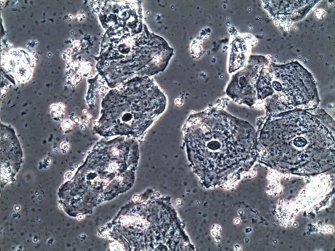
Oral Epithelial Cells – Phase Contrast
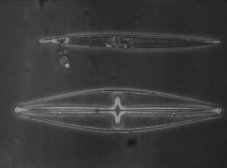
Diatom – Phase Contrast
Application – What is Dark Field Microscopy Used for
The dark field microscopy is mainly used to observe transparent specimens or unstained samples that have insufficient contrast in bright field imaging. Common samples include live aquatic organisms, diatom, bacteria, yeast, protozoa, chloroplast, mitochondria, histocyte, etc.
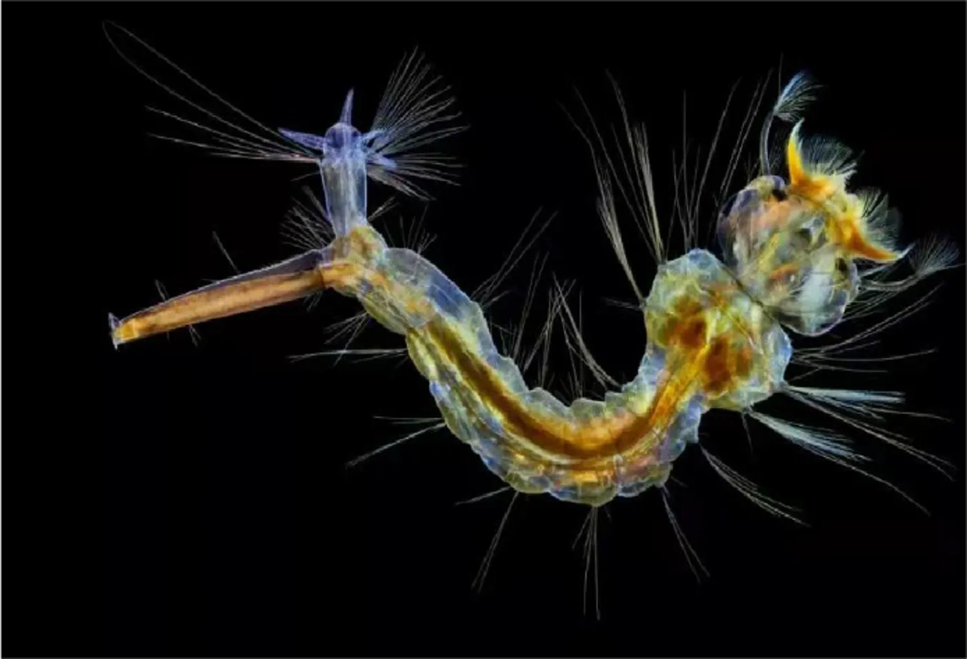
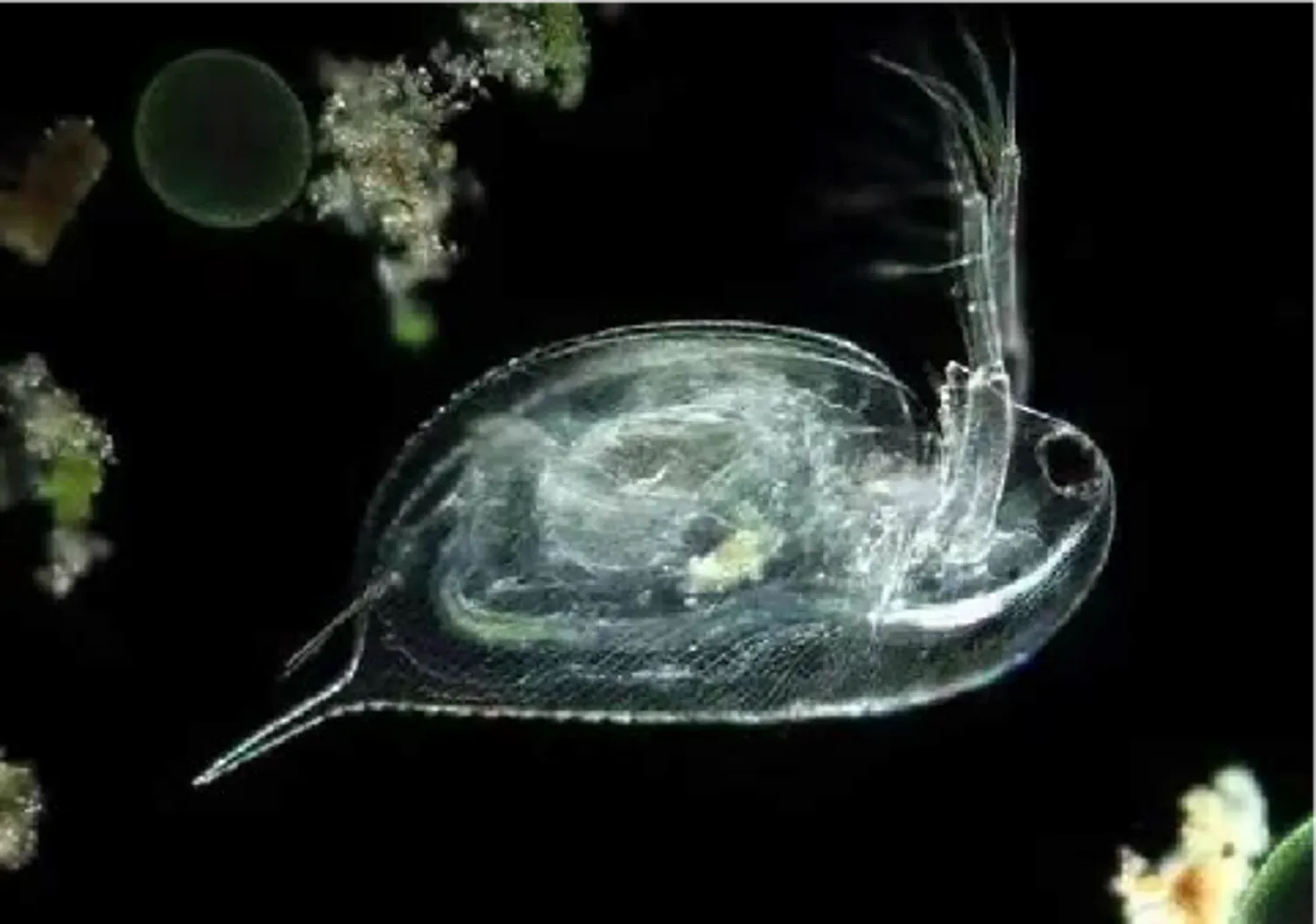
Living aquatic organisms
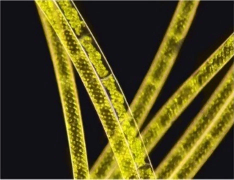
Spirogyra – Dark Field
The dark field microscopy can also be used to observe samples with smooth surface. In bright field microscopy, the surface details of such samples are vague because of the reflection halo. But in dark field, oblique illumination avoids strong reflected light, allowing the surface details to be clearly seen. Common applications include observing mineral, chemical crystal, gemstone inclusions, colloidal particles, dust, polymer with minor inclusions, ceramic thin sheets with cracks and inclusions, etc.
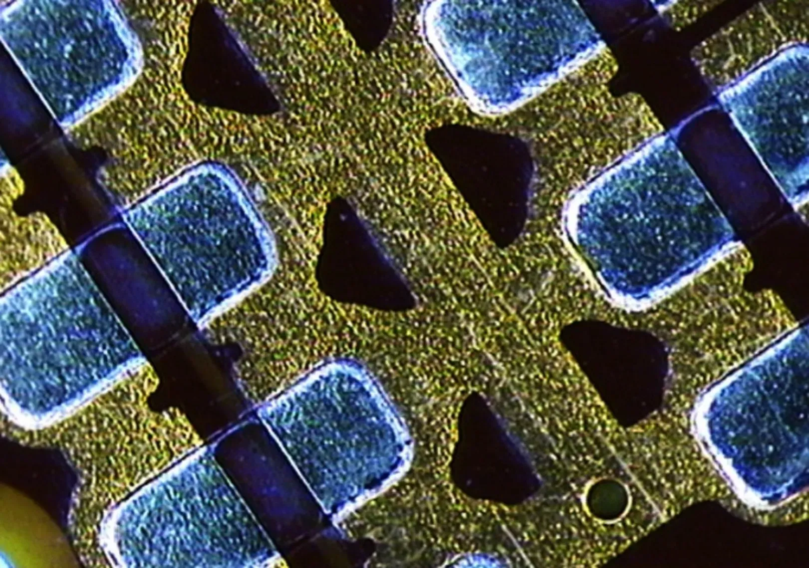
Wafer – Dark Field
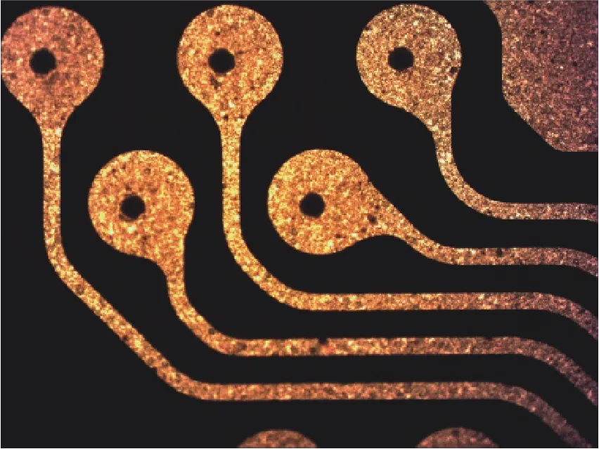
FPC – Dark Field
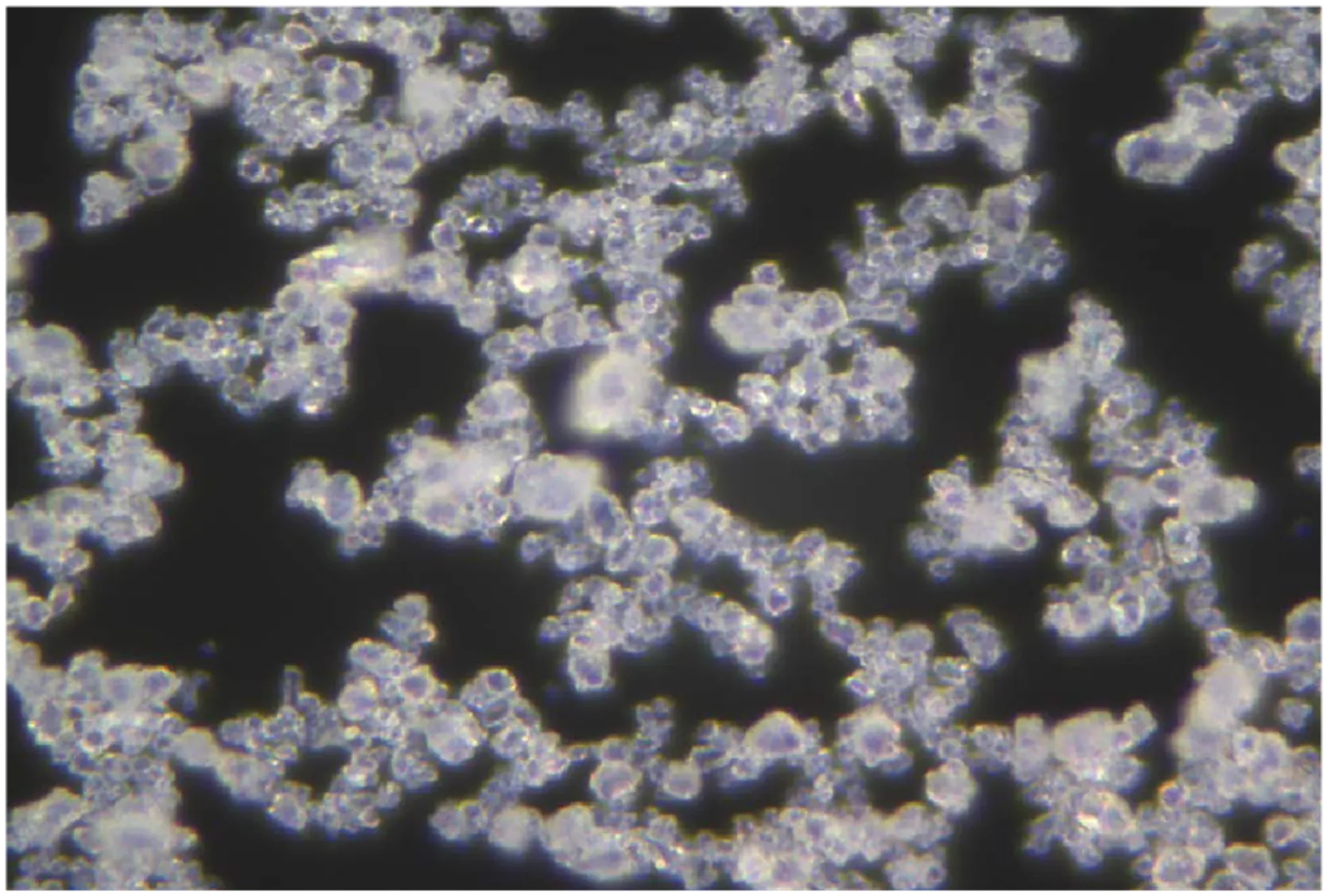
Crystal – Dark Field
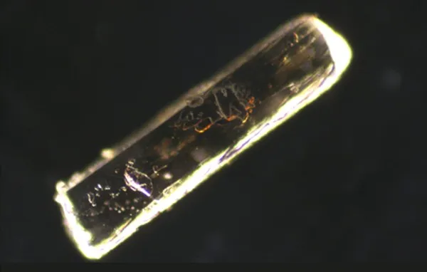
Sodium Glutamate Crystal – Dark Field
The dark field microscopy is very effective for observing as edges, boundaries, contours, refractive index gradient and features of the sample surface such as hairs, fibers, etc.
However, if there are complex and dense internal structure in the transparent sample, the dense structures will cause the scattered light to overlap, resulting in a blurry halo, which will affect the observation. Therefore, the dark field microscopy is not suitable for observing transparent samples with complex structure, such as thick tissue, dense bacterial cultures, etc. For such samples, phase contrast observation is a good choice.
Component – How to Achieve Dark Field Microscopy
Based on bright field microscope, to achieve dark field observation, you need some key components.
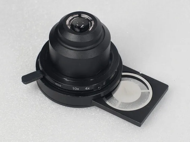
Dark field condenser
The dark field condenser is the key of dark field illumination. It generates a hollow light cone to illuminate the sample with oblique illumination. The condenser of some bright field microscopes can also achieve the function of a dark field condenser by installing a dark field slider. Note that since the realization of dark field requires the NA value of the objective to be lower that that of the condenser, when choosing objectives of different magnifications and NA values, a matching dark field condenser should be selected. Generally, a dry dark field condenser has an NA of 0.7-0.9 and is suitable for 4X-40X objectives, while an oil dark field condenser has an NA of 1.2-1.4 and is suitable for 60X-100X objectives. This is for reference only. It depends on the actual parameters of the objectives and the condensers.
Dark field objective
A compatible objective for dark field observation is required. Note that the NA value of the objective should be lower that that of the condenser.
High-intensity light source
Since dark field illumination requires blocking the central light, the light required for dark field observation is more than that for bright field observation. Therefore, a bright and high-intensity light source is needed.
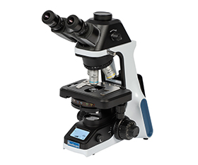
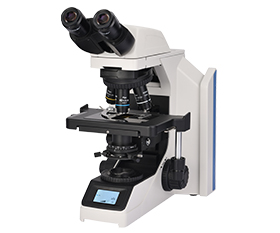
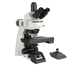
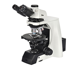
Dark field is a relatively easy observation method. Most compound microscopes can achieve dark field observation by adding a dark field kit.
Q&A
What is dark field microscopy used for?
Dark field is a lighting technique of oblique illumination that can achieve a bright sample image with a black background. It is used to observe transparent and low-contrast samples.
What kinds of samples are suitable for observation with a dark field microscope?
Thin and transparent samples, living cells and samples with smooth surfaces. The dark field microscope is suitable for observing edges, outlines and surface features.
What accessories are needed for dark field microscopy?
A dark field condenser or a dark field slider inserted into the existing condenser of the compound microscope, dark field objectives with a matching NA value to the condenser and a high-brightness light source.
Can samples for a dark field microscopy be unstained?
Yes. One of the main advantages of dark field microscopes is that the samples do not need to be stained. This not only avoids complex staining steps but also enables the observation of living samples.
What are the limitations of dark field microscopes?
The limitations include the need for high-intensity lighting, sensitivity to dust and impurities on the slides, unsuitability for observing thick and internally complex transparent samples, and the need to consider the matching of the NA values of the condenser and the objectives.
How to select a dark field condenser?
The NA value of the dark field condenser needs to be higher than that of the objectives. Generally, dry dark field condensers have an NA of 0.7-0.9 and are suitable for 4X-40X objectives, while oil dark field condensers have an NA of 1.2-1.4 and are suitable for 60X-100X objectives. This is for reference only. It depends on the actual parameters of the objectives and the condensers.
BestScope Dark Field Kit
Outfit
|
Dark Field Condenser |
Infinite Plan Objective 100X |
Infinite Plan Objective 100X with Iris Diaphragm |
Plan Objective 100X with Iris Diaphragm |
Achromatic 100X with Cone Diaphragm |
|
|
BDFA-D |
● |
|
|
|
|
|
BDFA-O |
● |
● |
○ |
|
|
|
BDFB-D |
● |
|
|
|
|
|
BDFB-O |
● |
|
|
○ |
● |
Note: ●Standard Outfit, ○Optional
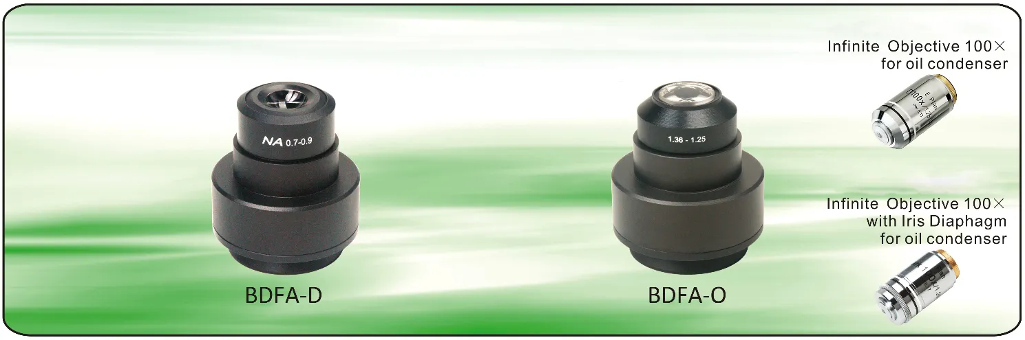
BDFA-D: NA 0.7-0.9 Dry Dark Field Condenser for Laboratory Microscope
BDFA-O: NA 1.25-1.36 Oil Dark Field Condenser for Laboratory Microscope
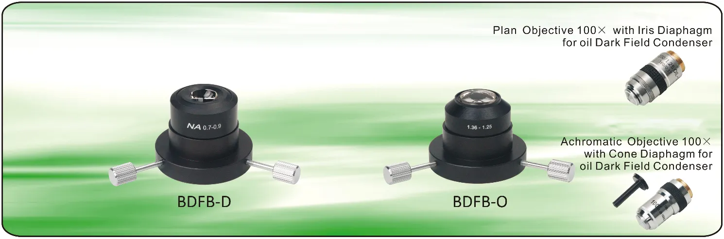
BDFB-D: NA 0.7-0.9 Dry Dark Field Condenser for Academic Microscope
BDFB-O: NA 1.25-1.36 Oil Dark Field Condenser for Academic Microscope