FISH Microscopy Instrument Solution
What’s FISH?
Fluorescence in situ hybridization, FISH, is a non-radioactive in situ hybridization technology developed on the basis of the radioactive in situ hybridization technology in the late 80s of the 20th century. At present, this technology has been widely used in different fields such as the research on the genomic structure of animals and plants, the analysis of fine structural variations of chromosomes, the analysis of viral infections, human prenatal diagnosis, tumor genetics and genome evolution research. It can be used for chromosome localization and gene mapping of genes or DNA fragments, detection of chromosomal number and structural abnormalities and research in hematological oncology and solid oncology, etc.
How to do FISH?
The basic principle of FISH is to use single-stranded nucleic acids directly or indirectly labeled with fluorescein as probes, and then hybridize the probes with the target DNA on chromosome or DNA fiber sections. According to the principle of base complementarity, if the two are homologous and complementary, a hybrid of the target DNA and the nucleic acid probe can be formed, which is a detectable hybrid double-stranded nucleic acid.
The indirect labeling method is to use nucleic acid probes labeled with non-fluorescent substances, such as biotin or digoxin and other labeled probes. And then fluorescent substances are brought in by affinity ligation or immune reactions to detect the presence of hybrids.
After hybridization, qualitative, quantitative or relative localization analysis of the target DNA can be conducted under the fluorescent microscope. Compared with the traditional radiolabeled in situ hybridization, fluorescence in situ hybridization has high speed, strong detection signal, high hybridization specificity and multiple staining. So, it has received widespread attention in different field.
Microscopy Instrument Solution of FISH
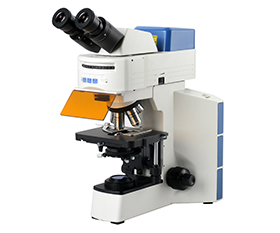
Fluorescence microscope
The general steps of FISH are probe denaturation, sample denaturation, hybridization, elution, hybridization signal amplification (applicable to probes labeled with biotin), counterstaining, sealing and observing FISH results under a fluorescence microscope.
The samples should be tested and observed as soon as possible after sealing. Each observation with a fluorescence microscope quenches a portion of the signal, so it is essential to observe and capture images quickly.
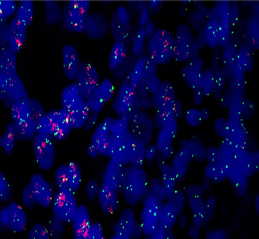
During observation, first find the cell division image in the field of view under the visible light source. And then turn on the fluorescence excitation illumination. The excitation wavelength of FITC is 490 nm. The cells will be stained red by PI, while the locations where the probes labeled by FITC were located shows green fluorescence.
Take the application of FISH in breast cancer detection as an example. First, confirm the cancer cell area on the stained section and observe the tissue cell structure under the 10X objective. Then scan the entire section with the 40X objective to observe whether there was heterogeneity in HER2 status. Finally, the FISH results of the cancer nucleus can be observed through a fluorescent filter under the 100X objective and the signal count and ratio can be calculated.
Recommended Fluorescence Microscope
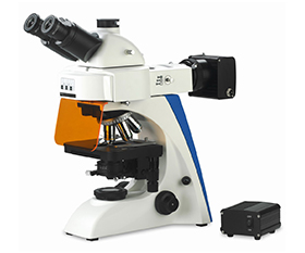
BS-2063F(LED) LED Fluorescence Microscope
BS-2063F2(LED) LED Fluorescence Microscope
BS-2064F Fluorescent Biological Microscope
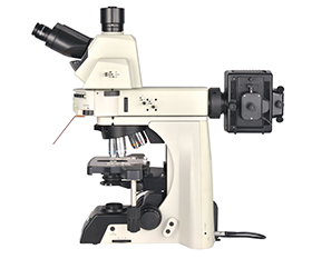
BS-2076F Research Fluorescent Biological Microscope
BS-2081F Research Fluorescent Biological Microscope
BS-2082F Research Fluorescent Biological Microscope
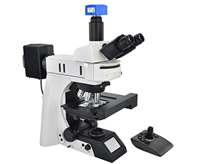
BS-2085F Motorized Fluorescent Biological Microscope
BS-2091F Inverted Fluorescent Biological Microscope
BS-2097 Research Inverted Microscope
BS-2098 Research Inverted Microscope
Confocal microscope
If possible, choose confocal microscopes to get better images. Compared with fluorescence microscopes, it improves the signal-noise ratio and has better resolution. Due to the limitation of optical diffraction, the imaging resolution of FISH probes is about 300 nm when using fluorescent microscope, so it will not be possible to accurately count two or more fluorescent probes in the nucleus when they are less than the imaging resolution. But confocal microscope can capture high resolution images. Also, multiple synaptic structural markers can be visualized simultaneously through fluorescence immunohistochemistry with confocal microscope. But the imaging speed is relatively slow, so it is not commonly used for diagnosis.
Recommended Confocal Microscope
BCF297 Laser Scanning Confocal Microscopy
Recommended Microscope Digital Camera
BUC5F Series C-mount USB3.0 CMOS Camera (HISPVP)
BUC5IB Series Cooled C-mount USB3.0 CMOS Camera
BUC5IC Series TE-Cooling M52/C-mount USB3.0 CMOS Camera
Fluorescent Filters for FISH
Specification
|
Item |
Specification |
|
|
Fluorescent Filters for FISH |
FISH-Green |
EX490/20nm, Dia25*5.0mm EM530/25nm, Dia25*3.5mm DM505nm, 36*25.7-1.05mm |
|
FISH-Blue |
EX410/20nm, Dia25*5.0mm EM480/30nm, Dia25*3.5mm DM455nm, 36*25.7*36-1.05mm |
|
|
FISH-Orange |
EX540/20nm, Dia25*5.0mm EM590/25nm, Dia25*3.5mm DM555nm, 36*25.7-1.05mm |
|
|
FISH-Red |
EX580/20nm, Dia25*5.0mm EM630/25nm, Dia25*3.5mm DM605nm, 36*25.7-1.05mm |